Inferior medullary velum (Latin: velum medullare inferius)
Also called the posterior medullary velum
The inferior medullary velum is:
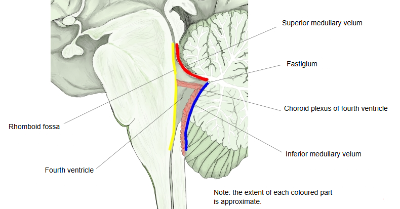
It is a thin sheet of white matter stretched between the nodule of the cerebellum and the peduncles of the flocculus, and forms part of the lower roof of the fourth ventricle (whose floor is the rhomboid fossa).
The walls of the fourth ventricle are as follows.
The fourth ventricle is tent-shaped, between the flat rhomboid fossa (its floor) and the superior medullary velum and inferior medullary velum, which run from the edges of the fossa towards the cerebellum and meet at the peak (the fastigium).
The lower half of the back wall is formed by the inferior medullary velum and the tela choroidea, bounded on each side by the inferior cerebellar peduncles. The ventricle widens to each side into a lateral recess, which ends in the lateral aperture (foramen of Luschka). The vessel-rich choroid plexus of the fourth ventricle is attached to the lower roof, and its lateral ends stick out through the lateral apertures (sometimes called Bochdalek's flower basket). In the midline of the lower roof, just above the medulla oblongata, is the median aperture (foramen of Magendie). Through these openings cerebrospinal fluid leaves the ventricles for the subarachnoid space.
Some textbooks divide the roof of the fourth ventricle into an upper (front) half and a lower (back) half, made up as follows.
- Upper half : the superior medullary velum and the superior cerebellar peduncles (brachia conjunctiva)
- Lower half : the inferior medullary velum and the tela choroidea of the fourth ventricle (with the choroid plexus of the fourth ventricle)
Note: some sources also count the nodule of the cerebellar vermis in the lower half.
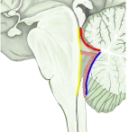 Fourth ventricle (median section)
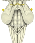 Rhomboid fossa
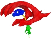 Ventricles: left side (coloured)
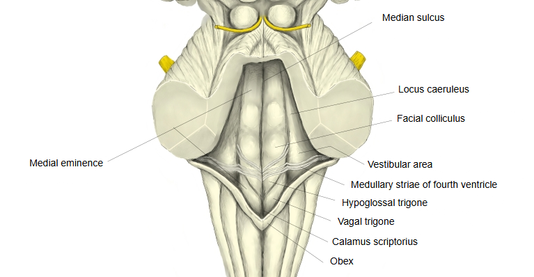
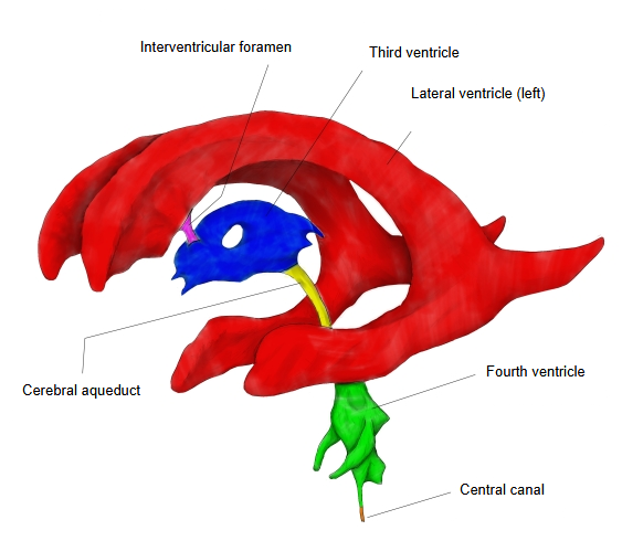
The inferior medullary velum (posterior medullary velum) is a thin layer of white matter continuing from the white centre of the cerebellum, above and on each side of the nodule; it forms the lower back part of the roof of the fourth ventricle. It is roughly half-moon-shaped: its convex edge is continuous with the white matter of the cerebellum, while its thin concave edge looks free but in fact continues into the ependymal lining of the ventricle (with the tela choroidea), which extends down to the taeniae of the fourth ventricle.
Reference websites
· Inferior medullary velum and Fourth ventricle, Wikipedia.
· Scheme of the roof of the fourth ventricle (Gray's Anatomy, fig. 708) (theodora.com)

· The same figure on Wikimedia Commons

Related terms
- Choroid plexus
- Third ventricle
- Inferior horn of lateral ventricle
- Vein of lateral recess of fourth ventricle
- Posterior inferior cerebellar artery
- Brain
References and sources
- Further reading: Inferior medullary velum, Wikipedia.
- Further reading: Fourth ventricle, Wikipedia.
- Federative International Programme for Anatomical Terminology (FIPAT). Terminologia Anatomica, 2nd ed. (TA2), 2019.