Rhomboid fossa (Latin: fossa rhomboidea)
Also called the floor of the fourth ventricle
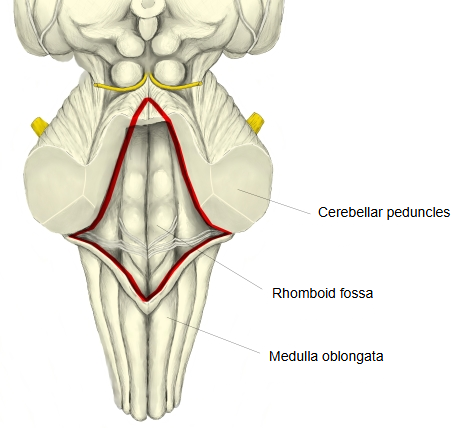
· It is formed by the back of the pons (upper half) and of the upper medulla oblongata (lower half); the cavity of the fourth ventricle lies between the brainstem in front and the cerebellum behind.
The fourth ventricle is divided into two main parts:
ventricle
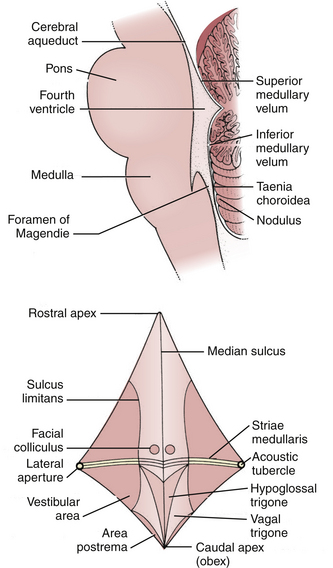
- Rhomboid fossa : the diamond-shaped, shallow floor of the fourth ventricle.
- Roof of fourth ventricle : the roof of the fourth ventricle, facing and covered by the cerebellum.
It lies on the back of the medulla oblongata and the pons; at the sides it is joined to the cerebellum by the cerebellar peduncles.
→ its edges are formed by the superior cerebellar peduncles above and the inferior cerebellar peduncles below; the middle cerebellar peduncles join the cerebellum at the lateral angles.
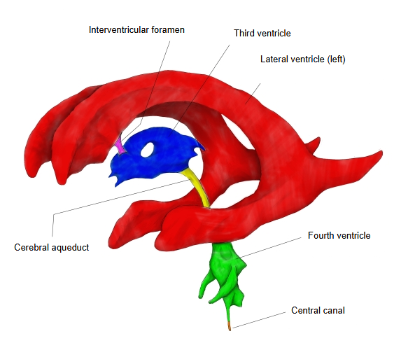
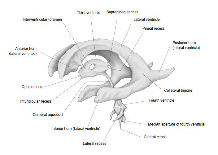
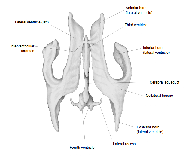
· It is about 3 cm long and 2 cm wide (sources differ on the width). Above, the fourth ventricle continues into the cerebral aqueduct; below, into the central canal of the spinal cord. The lateral recesses extend sideways from the widest part.
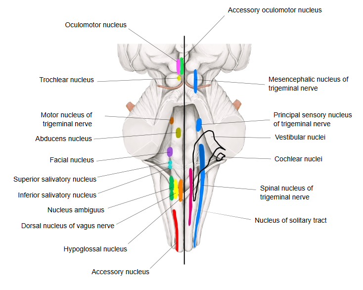
Many nerve nuclei lie beneath the rhomboid fossa: the nuclei of cranial nerves V–XII, so the fossa is a useful map of the brainstem.
· Abducens nucleus: under the facial colliculus (nucleus of the abducens nerve).
· Vestibular nuclei: under the vestibular area (nuclei of the vestibular nerve).
· Nucleus of locus caeruleus: under the locus caeruleus.
· Hypoglossal nucleus: under the hypoglossal trigone (nucleus of the hypoglossal nerve).
· Dorsal nucleus of vagus nerve: under the vagal trigone (a nucleus of the vagus nerve).
Surface landmarks
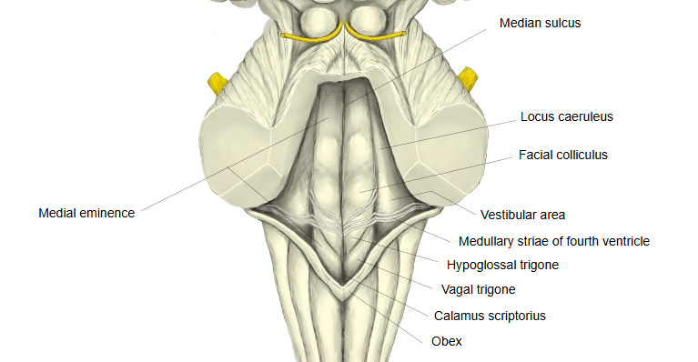
· Median sulcus: the groove running down the midline of the floor, dividing it into right and left halves; below, it is continued by the posterior median sulcus of the medulla oblongata and the spinal cord.
· Medial eminence: the raised strip on each side of the median sulcus.
· Sulcus limitans: a groove lateral to the medial eminence; motor nuclei lie medial to it, sensory nuclei lateral to it.
· Facial colliculus: a swelling of the medial eminence in the upper (pontine) part, made by fibres of the facial nerve looping round the abducens nucleus.
· Medullary striae of fourth ventricle: thin, thread-like fibre bundles running across the floor at its widest part, at the border between pons and medulla.
· Vestibular area: the slightly raised area lateral to the sulcus limitans, the groove that runs lateral to the medial eminence; it lies over the vestibular nuclei.
· Locus caeruleus: a dark bluish-grey area at the upper end of the sulcus limitans, in the upper (pontine) part of the floor above the vestibular area; the nucleus of the locus caeruleus lies beneath it.
· Hypoglossal trigone: the raised area in the medial part of the floor below the medullary striae, over the hypoglossal nucleus.
· Vagal trigone (also called the ala cinerea): a small triangular area lateral to the hypoglossal trigone, over the dorsal nucleus of the vagus nerve.
· Calamus scriptorius: the lower tip of the rhomboid fossa, shaped like the nib of a pen.
· Obex: the small fold at this tip, formed by the taenia of the fourth ventricle, where the fourth ventricle narrows into the central canal.
 Brainstem, posterior view (cerebellum removed)
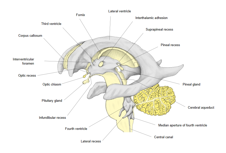
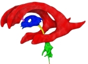 Ventricles: left side (coloured)
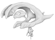 Ventricles: left side
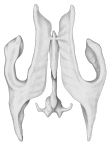 Ventricles: from above
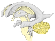 Ventricles and surroundings
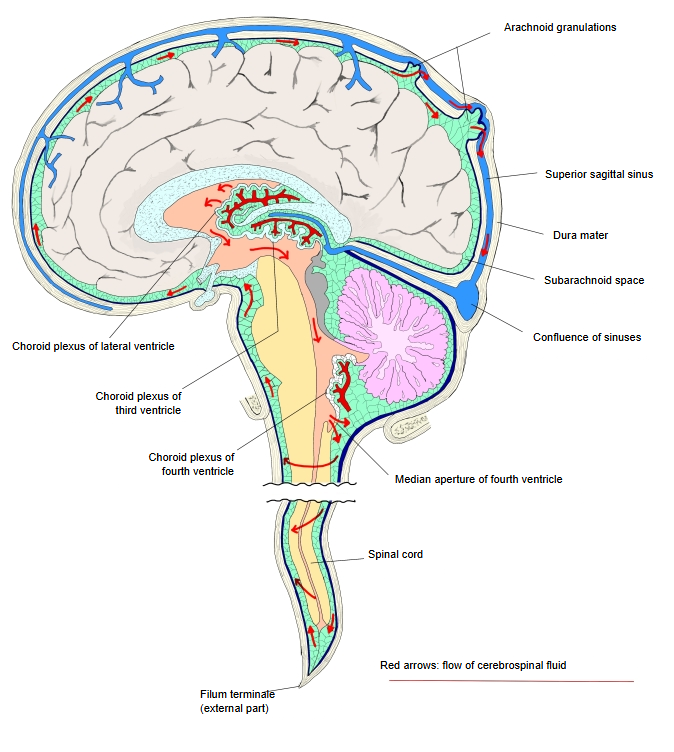
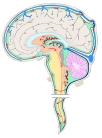 Flow of cerebrospinal fluid
Reference websites
[Wikipedia]
· Rhomboid fossa, Wikipedia.
· Fourth ventricle, Wikipedia.
[Websites with illustrations]
· Rhomboid fossa (figure) (Kenhub)

· Fourth ventricle in sagittal section and its floor, with landmarks (figure) (Neupsy Key)
Related terms
- Pia mater
- Cerebral ventricles
- Inferior medullary velum
- Third ventricle
- Choroid plexus
- Brain
- Facial nerve
- Vagus nerve
- Hypoglossal nerve
References and sources
- Further reading: Rhomboid fossa, Wikipedia.
- Further reading: Fourth ventricle, Wikipedia.
- Federative International Programme for Anatomical Terminology (FIPAT). Terminologia Anatomica, 2nd ed. (TA2), 2019.