Cystohepatic triangle (Latin: trigonum cystohepaticum)
Also called Calot's triangle or the hepatocystic triangle
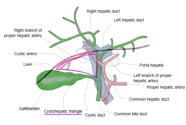
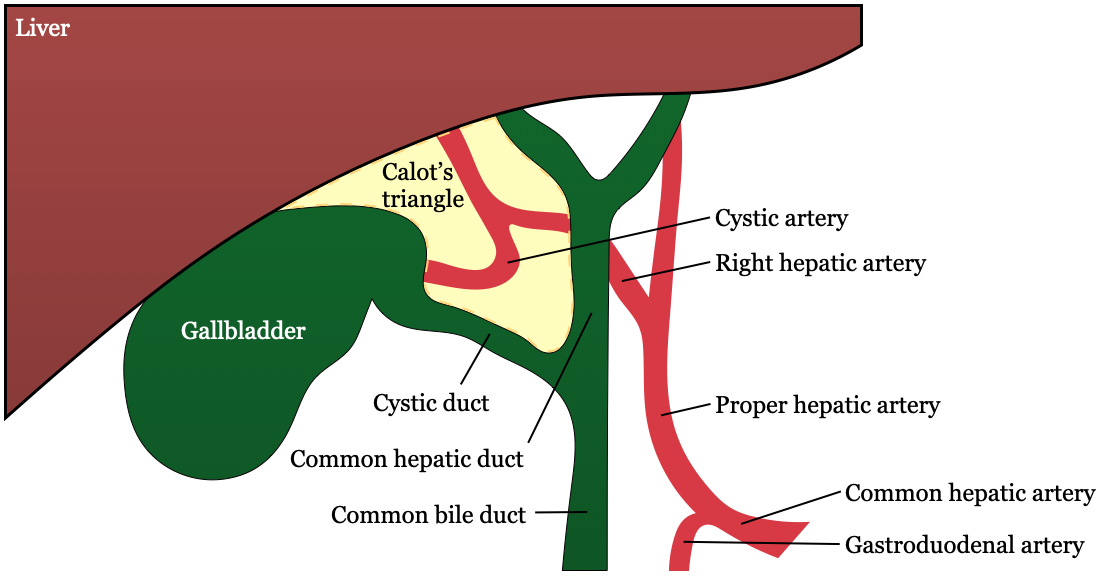
It lies near the neck of the gallbladder, below the porta hepatis. Its borders are the cystic duct on the gallbladder side, the common hepatic duct on the midline side, and the underside of the liver above. It usually contains the cystic artery, often the right branch of the proper hepatic artery, and the cystic lymph node. In the original description by Jean-François Calot (1891) the upper border was the cystic artery, not the liver, so the triangle was smaller; sources differ on which border is used, and the larger form is often called the hepatocystic triangle. It is a key landmark in gallbladder removal (cholecystectomy): the cystic duct and cystic artery are identified here before they are divided, above their junction with the common bile duct.
Reference websites
[Wikipedia]
· Cystohepatic triangle, Wikipedia.
[Websites with illustrations]
· Calot's triangle: borders and contents (TeachMeAnatomy)
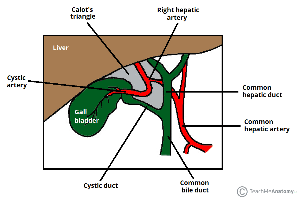
· Calot's triangle (Surgery Student)
Related terms
- Cystic duct
- Common hepatic duct
- Neck of gallbladder
- Gallbladder
- Cystic artery
- Common bile duct
- Porta hepatis
References and sources
- Further reading: Cystohepatic triangle, Wikipedia.
- Federative International Programme for Anatomical Terminology (FIPAT). Terminologia Anatomica, 2nd ed. (TA2), 2019.