Gallbladder (Latin: vesica biliaris; vesica fellea)
- Overview
- Divisions
- Structure
- Functions
- Vessels and nerves (arteries, veins, nerves, lymphatics)
- Related terms
- References
Overview
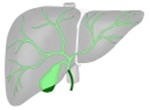 Liver, bile ducts and gallbladder
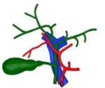 Arteries, veins and bile ducts near the portal vein
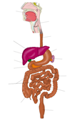 Digestive system
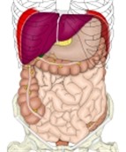 Organs in the abdominal cavity
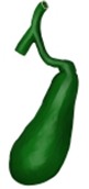 The gallbladder, whole view
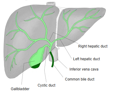
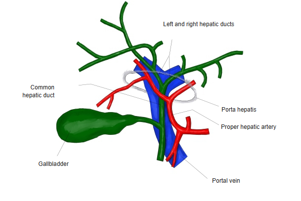
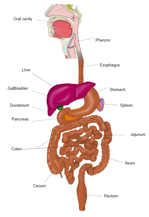
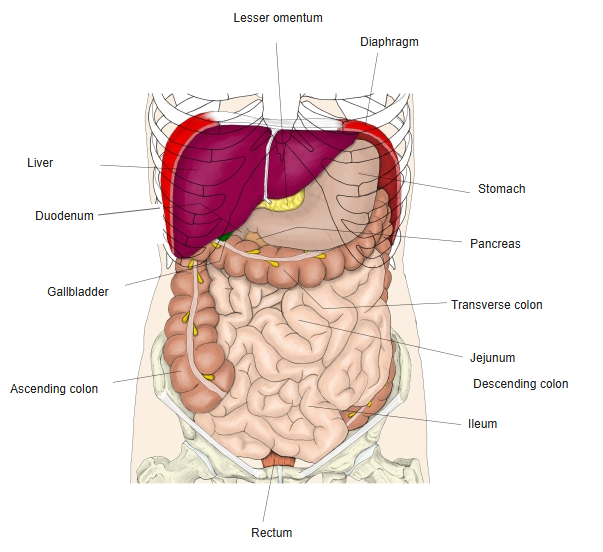
 Position of the gallbladder
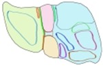 Names on the visceral surface of the liver
 Visceral surface of the liver 1
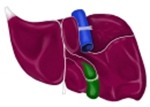 Visceral surface of the liver 2
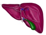 Liver (posterior surface)
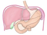 The gallbladder and other organs 1
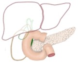 The gallbladder and other organs 2
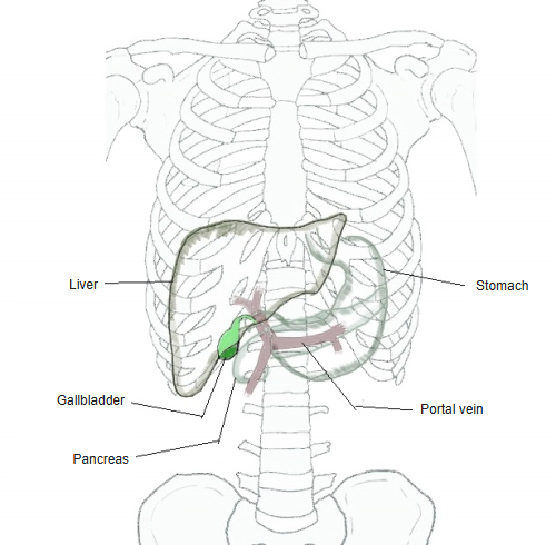
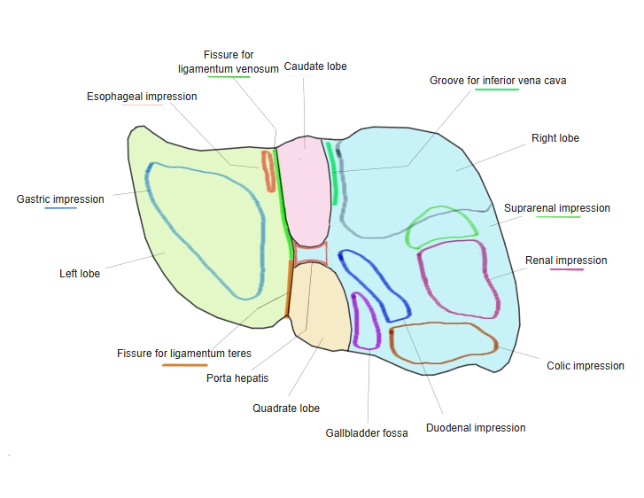
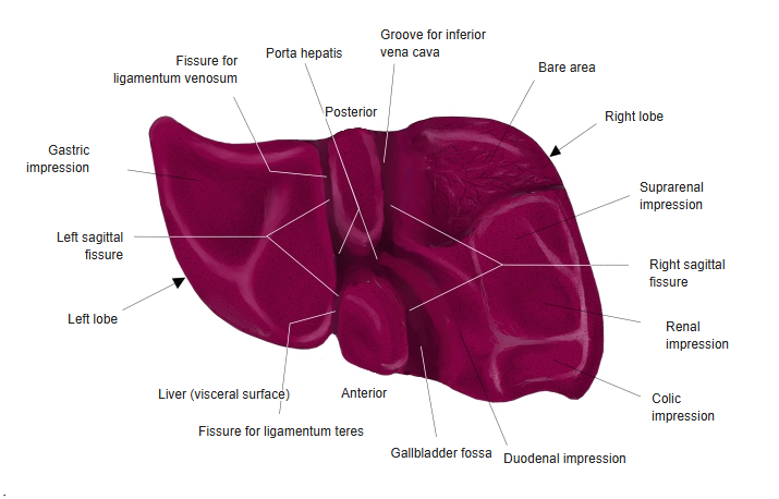
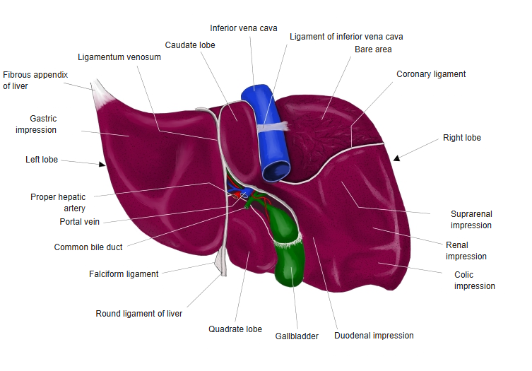
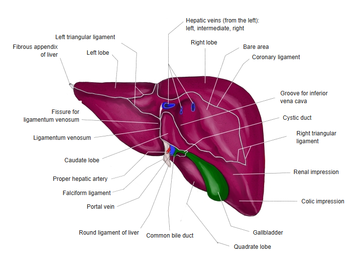
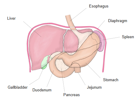
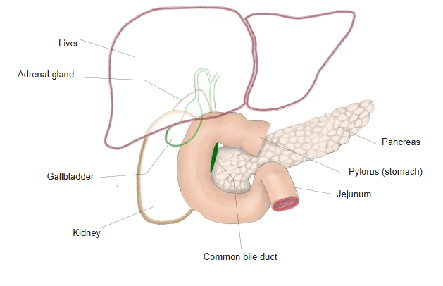
The size of the gallbladder changes greatly with the amount of bile stored inside. While fasting, bile is stored in the gallbladder and it swells; after a meal, especially one containing fat, a hormone (cholecystokinin) is secreted, the gallbladder contracts and sends bile into the duodenum, and so it gradually becomes smaller.
Cholecystokinin is secreted by the endocrine "I cells" of the upper small intestine (especially the duodenum and the upper jejunum).
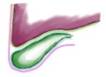 Upper and lower surfaces of the gallbladder
 The gallbladder and other organs
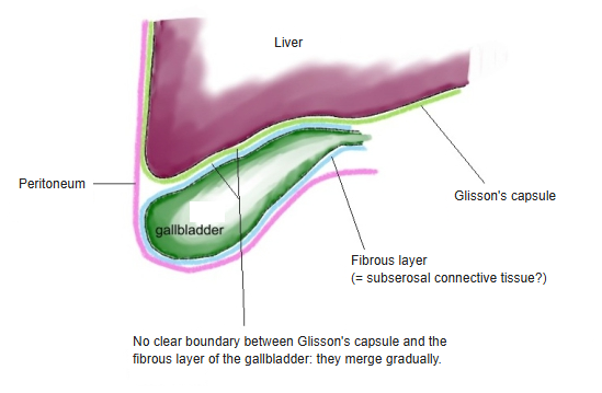
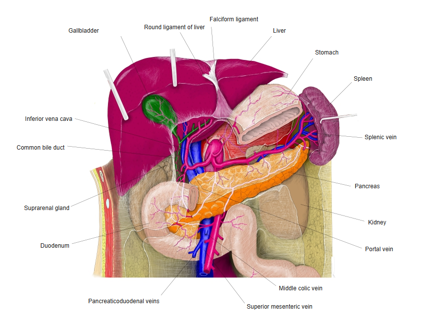
[Some figures]
1. Volume ratio of the gallbladder to the liver: about 1 : 25–50.
2. Thickness of the gallbladder wall: normally about 1–3 mm.
3. Is the gallbladder green? The tissue of the gallbladder itself is not green but greyish-white to pale pink; it often looks green because the bile stored inside is green to yellowish brown.
Divisions
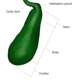
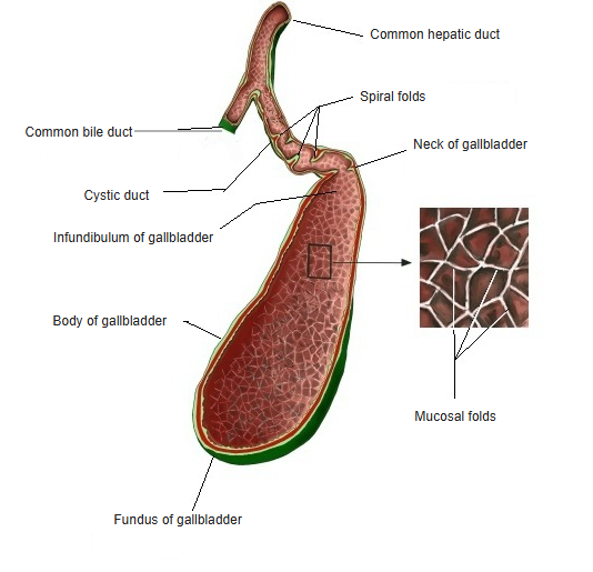
On this site the cystic duct is treated as part of the bile ducts, and the gallbladder is divided into 3 parts: the neck, the body and the fundus.
Some textbooks, however, count the cystic duct as part of the gallbladder. This may be revised if needed.
Each part is briefly described below, but there are no clear borders between the parts.
the
gallbladder
- Neck : the part continuing from the cystic duct at the posterior upper end.
- Hartmann's pouch : a slightly widened part where the body passes into the neck.
- Body : the large middle part lying between the neck and the fundus.
- Fundus : the rounded anterior lower end, facing downwards.
- There are no clear borders between the parts.
Structure
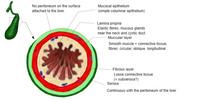
Histologically, the gallbladder wall has 3 layers (mucosa, muscular layer, serosa).
The gallbladder has the same basic structure as other hollow organs, but is characterised by having no distinct submucosa and no muscularis mucosae of smooth muscle.
| 1 | Mucosa | a | Epithelium | Simple columnar epithelium (tall cells with a thin brush border). Joined to the lamina propria through a thin basement membrane. The epithelial cells absorb water and inorganic salts from the bile and concentrate it 6–10 times. The mucus they secrete protects the wall from the concentrated bile and keeps the bile flowing smoothly. |
| b | Lamina propria | Elastic fibres are present; from the neck to the cystic duct there are small tubulo-alveolar mucous glands. | ||
| 2 | Muscular layer | Smooth muscle mixed with connective tissue fibres. [Course of the smooth muscle] · Circular fibres: the main layer, especially many in the neck. · Oblique / longitudinal fibres: many in the body / fundus. | ||
| 3 | Serosa | a | Fibrous layer | Loose connective tissue enclosing the muscular layer (= subserosal connective tissue?). |
| b | Serosa | Peritoneum (covers the lower surface of the gallbladder). | ||
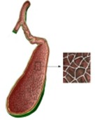 Section of the gallbladder 1
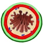 Section of the gallbladder 2
Functions
■ Storage of bile ■
It temporarily stores the bile made continuously by the liver (0.6–1 L a day).
■ Concentration of bile ■
The epithelial cells lining the gallbladder reabsorb water and electrolytes and concentrate the bile 5–10 times.
⇒ Bile is stored efficiently in a limited space.
■ Release of bile ■
When food (especially fat) enters the duodenum, a hormone called cholecystokinin (CCK) is secreted and the gallbladder contracts.
At the same time the sphincter of Oddi relaxes, and bile is released through the common bile duct into the duodenum.
Vessels and nerves
[Arteries]
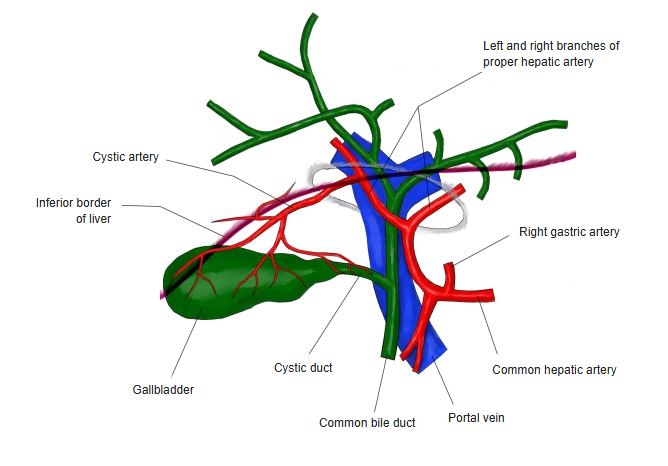
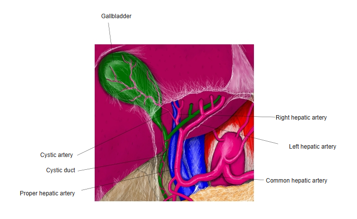
The cystic artery arises from the proper hepatic artery (usually from its right branch), divides into 2 branches and supplies the gallbladder from the side.
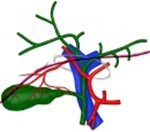 Proper hepatic artery and its branches
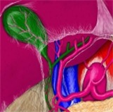 Cystic artery
[Veins]
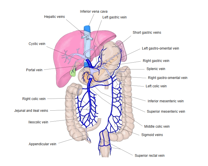
The cystic veins, which collect the blood from the gallbladder, drain into the portal vein.
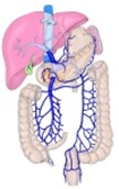 Digestive system and veins
[Nerves]
■ Sympathetic fibres ■
Origin: thoracic spinal cord (T5–T9).
Route: celiac plexus → hepatic plexus → gallbladder.
Action: inhibits contraction of the gallbladder and contracts the sphincter of Oddi.
Types: contain both efferent (motor) and afferent (pain) fibres.
■ Parasympathetic fibres ■
Origin: the vagus nerve.
Route: anterior vagal trunk → through the hepatic plexus to the gallbladder.
Action: contracts the gallbladder and relaxes the sphincter of Oddi.
Types: mainly efferent (motor) fibres.
■ Visceral afferent fibres ■
They carry visceral sensations such as pain and discomfort (afferent nerve fibres).
The pain felt in the right shoulder and right upper abdomen during a gallstone attack (referred pain) is carried by these afferent fibres.
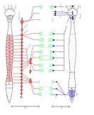 Distribution of the autonomic nervous system
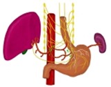 Vagus nerve (thorax and abdomen)
 Autonomic nerves and viscera
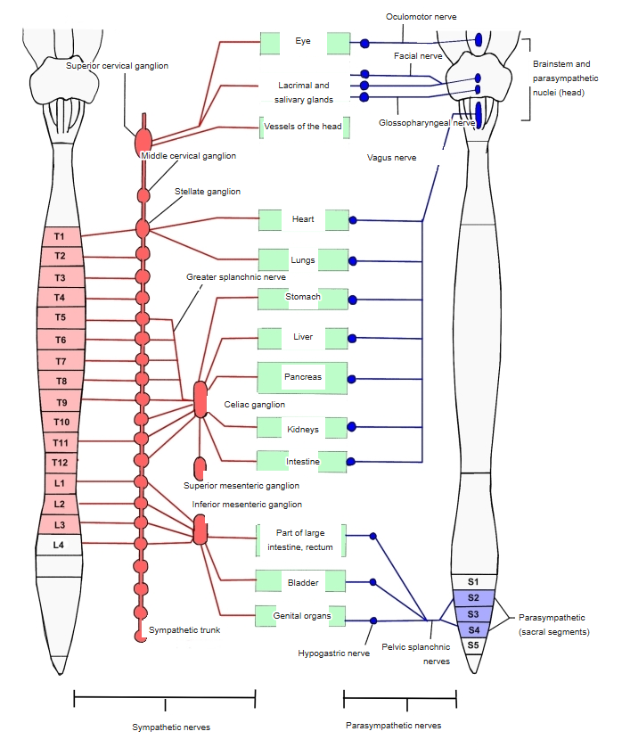
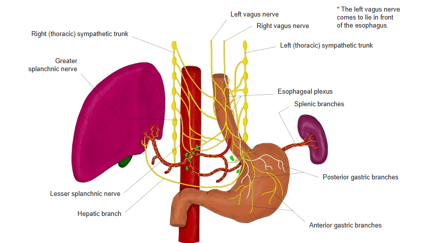
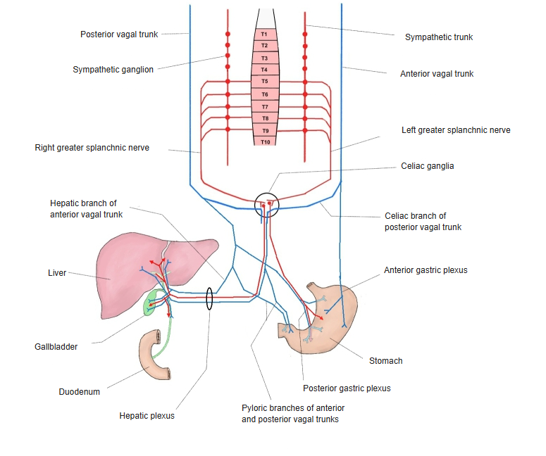
[Lymphatics]
Lymph vessels are present throughout the gallbladder wall, in the mucosa, the muscular layer and the subserosal tissue, and help remove waste and foreign matter arising in the wall.
Lymph drainage from the gallbladder
Lymph from the gallbladder passes mainly through the following lymph nodes:
cystic lymph node (Lund's node)
→ hepatic nodes
→ celiac nodes.
Related terms
Main anatomical terms related to the gallbladder, in alphabetical order.
| B | Body of gallbladder |
| C | Common hepatic duct, Cystic duct |
| F | Fundus of gallbladder |
| I | Infundibulum of gallbladder |
| N | Neck of gallbladder |
| S | Spiral folds |
References and sources
- Further reading: Gallbladder, Wikipedia.