Articular cavity (Latin: cavitas articularis)
Also called Joint cavity
 Model of a synovial joint
 Hip joint (coronal section)
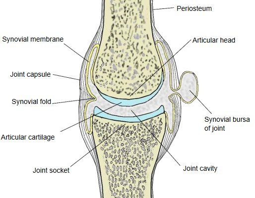
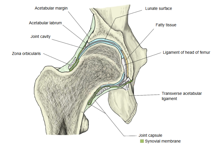
The whole joint is wrapped in the joint capsule, which is continuous with the periosteum of the bones, so the gap between the bones forms a closed joint cavity. The cavity is a narrow, slit-like space that contains synovial fluid.
Its walls are the synovial membrane (the inner layer of the joint capsule) and the articular cartilage on the ends of the bones. It normally holds only a thin film of synovial fluid, so it is a potential space rather than an open cavity.
· Only synovial joints have a joint cavity; fibrous and cartilaginous joints do not.
· An articular disc divides the cavity completely into two compartments, as in the temporomandibular joint; a meniscus divides it only partly, as in the knee joint (see the lateral meniscus).
· After injury or inflammation, excess fluid or blood may collect in it (joint effusion).
Reference websites
[Wikipedia]
· Synovial joint, Wikipedia.
· Synovial fluid, Wikipedia.
[Websites with illustrations]
· Synovial joint (diagram) (Wikimedia Commons)

· Synovial joint anatomy (TeachMeAnatomy)
Related terms
- Synovial membrane
- Synovial fold
- Hip joint
- Knee joint
- Temporomandibular joint
- Lateral meniscus
- Plane joint
References and sources
- Further reading: Synovial joint, Wikipedia.
- Further reading: Synovial fluid, Wikipedia.
- Federative International Programme for Anatomical Terminology (FIPAT). Terminologia Anatomica, 2nd ed. (TA2), 2019.