Synovial membrane (Latin: membrana synovialis)
Also called: synovial layer
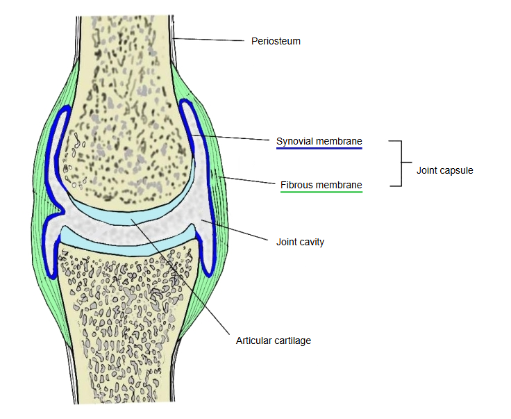
The membrane that forms the inner layer of the joint capsule; it consists of two layers. It is rich in blood vessels, produces synovial fluid, which it releases into the joint cavity, and through it supplies nutrients to the articular cartilage.
capsule
- Fibrous membrane : (fibrous capsule): the outer layer; a tough membrane rich in collagen fibres
- Synovial membrane : the inner layer; it consists of two layers
The diagram below sums up several sources, which are described after it.
membrane
- Intima (lining layer) : the layer facing the joint cavity, made of synovial cells: cells like fibroblasts, together with macrophages and a few lymphocytes
- Subintima : contains elastic fibres, blood vessels and nerves
The surface facing the joint cavity is covered by flat cells, and outside them lies connective tissue containing elastic fibres. The synovial membrane is rich in blood vessels and secretes the clear, viscous synovial fluid, which reduces friction between the joint surfaces during movement. It also projects into the joint cavity as synovial folds; folds containing much fat are called adipose folds (fat pads), and very small projections are called synovial villi. In large joints the synovial membrane may push out through the fibrous membrane to form a pouch containing synovial fluid, a synovial bursa of the joint; most of these lie under muscles or tendons and help them move smoothly. A part of the capsule may also bulge out as a pouch, called a sacciform recess (as in the elbow region).
The inner layer of the joint capsule is soft, loose connective tissue, called the synovial membrane (synovial layer). Many small synovial villi project from it, and synovial folds containing masses of fat tissue may fill the dead spaces in the joint. The fibrous membrane is thickened in places to strengthen the joint; ligaments are especially developed parts of it, or form separately from it. The synovial membrane consists of the surface synovial cells and a lamina propria containing elastic fibres, blood vessels and nerves. Its rich blood supply is directly related to the activity of the joint: joints that move a lot are generally richer in vessels than those that move little. Long-term irritation of the synovial membrane increases the production of synovial fluid, and fluid collects in the joint cavity (a joint effusion); the membrane may then bulge out through a weak part of the fibrous layer and form a synovial cyst.
The synovial membrane is very thin, and unlike an epithelium its cells do not sit side by side on a basement membrane: within its connective tissue lie fibroblast-like synovial cells, macrophages and a few lymphocytes. The membrane itself forms folds that help the joint move smoothly. The synovial cells produce the synovial fluid, which is rich in hyaluronan (hyaluronic acid) and the lubricating glycoprotein lubricin, and which nourishes the cartilage. Articular cartilage has no blood vessels, so its chondrocytes depend on the synovial fluid for nutrients; for this reason the synovial membrane is richly supplied with blood vessels. It lines all inner surfaces of the joint except the articular cartilage (and the menisci and discs), attaching at the margins of the cartilage.
Reference websites
[Wikipedia]
· Synovial membrane, Wikipedia.
· Synovial joint, Wikipedia.
Related terms
| 1 | Synovia (= synovial fluid) | the fluid in the joint cavity |
| 2 | Synovial fold | a fold of the membrane projecting into the joint cavity |
| 3 | Adipose folds | plicae adiposae: synovial folds containing much fat, such as the infrapatellar fat pad of the knee joint |
| 4 | Synovial villi | very small finger-like projections of the membrane |
| 5 | Synovial bursa of joint | a pouch of the membrane pushed out through the fibrous membrane |
| 6 | Sacciform recess | a pouch-like outpouching of the capsule |
Related terms
References and sources
- Further reading: Synovial membrane, Wikipedia.
- Further reading: Synovial joint, Wikipedia.
- Federative International Programme for Anatomical Terminology (FIPAT). Terminologia Anatomica, 2nd ed. (TA2), 2019.