Knee joint (Latin: articulatio genus)
Overview
The joint of the knee. It has the largest articular surfaces of any joint in the body.
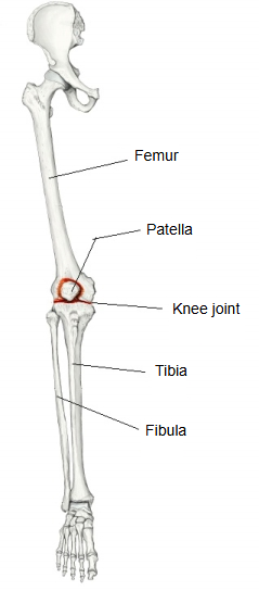
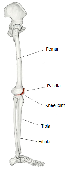
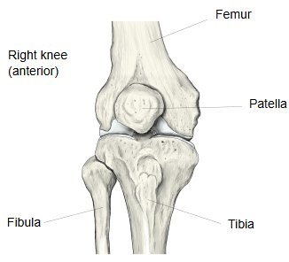
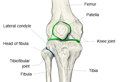
[Bones / type of joint]
Three bones form the knee joint: the femur, the tibia and the patella. The fibula takes no part in it. The joint is described below in two parts: the articulation between the femur and the tibia, and the articulation between the femur and the patella.
Femur and tibia
· This part is also called the femorotibial joint.
Femur and patella
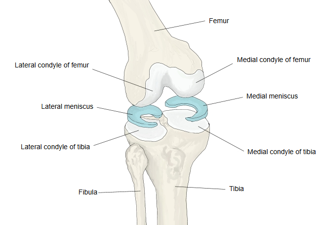
· This part is also called the patellofemoral joint.
- Tibia : medial and lateral condyles of each bone (hinge joint)
- Patella : patellar surface of the femur and posterior surface of the patella (saddle joint)
Joint capsule
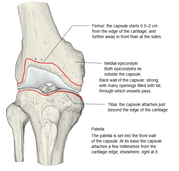
On the femur, the joint capsule is attached about 0.5–2 cm away from the edge of the articular cartilage, farther away in front than at the sides and behind. Both epicondyles of the femur lie outside the capsule. On the tibia it is attached close to the edge of the cartilage. The patella is set into the front wall of the capsule: the capsule is attached a few millimetres from the cartilage at the base of the patella and close to the cartilage along its other borders. The posterior wall is strong; it has many openings for blood vessels, and fatty tissue fills the spaces between them.
Joint cavity
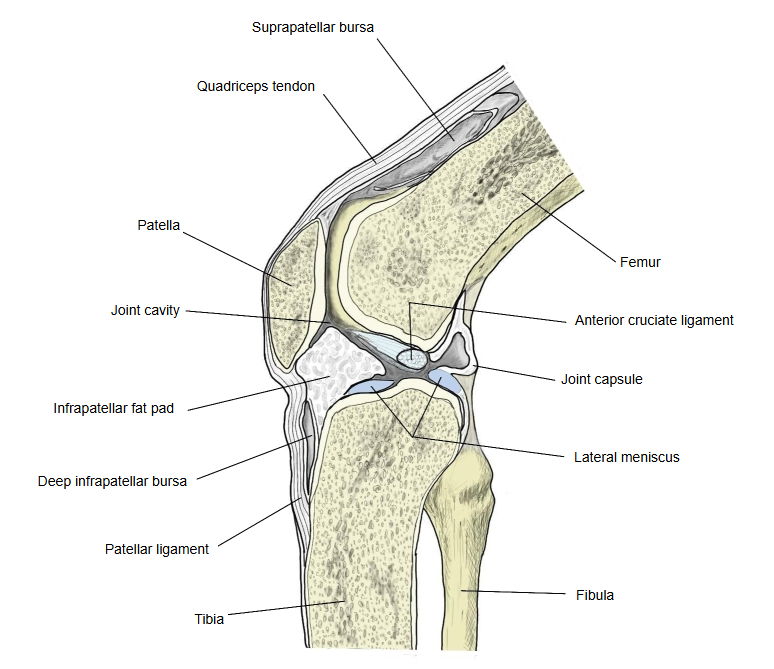
The joint cavity of the knee is the largest and the most complicated of all the joints. Followed from its upper posterior part, the synovial membrane passes over the cruciate ligaments and down to the tibia. Between the tibia and the patella the synovial membrane covers the infrapatellar fat pad, a broad mass of fat that bulges into the cavity. Two folds run onto the fat pad from the sides of the patella: the alar folds. From the middle of the fat pad a cord made only of synovial membrane crosses the cavity to the anterior cruciate ligament: the infrapatellar synovial fold. Its thickness varies, and it is often very thin.
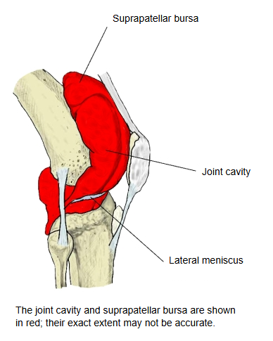
Synovial bursae
There are many synovial bursae around the knee joint. The table lists the main ones; it does not include every bursa around the knee, and some names in the table differ from the names in the illustrations.
| Name | Remarks | Illustration | |
|---|---|---|---|
| 1 | Suprapatellar bursa | Above the patella, behind the quadriceps tendon; it usually opens into the joint cavity. | 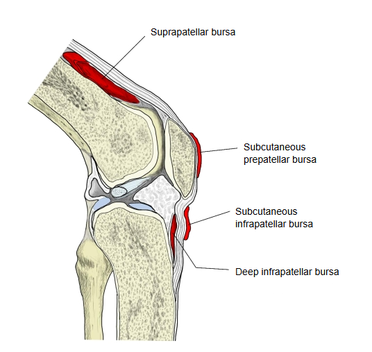 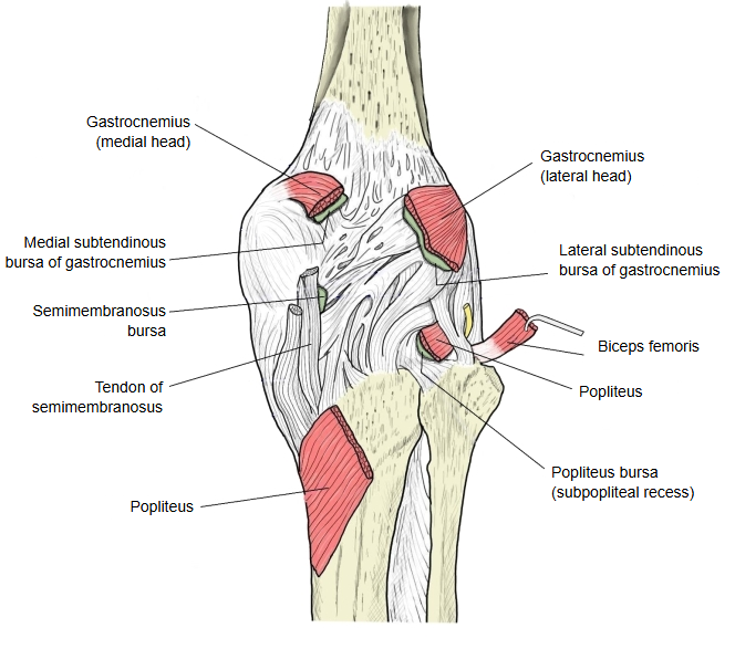 |
| 2 | Popliteus bursa (subpopliteal recess) | ||
| 3 | Semimembranosus bursa, tibial (medial) part | ||
| 4 | Semimembranosus bursa, fibular part | ||
| 5 | Bursa of the medial head of gastrocnemius | ||
| 6 | Gastrocnemius–semimembranosus bursa | ||
| 7 | Subcutaneous prepatellar bursa | ||
| 8 | Subfascial prepatellar bursa | ||
| 9 | Subtendinous prepatellar bursa | ||
| 10 | Subcutaneous infrapatellar bursa | ||
| 11 | Deep infrapatellar bursa | ||
| 12 | Subcutaneous bursa of the tibial tuberosity |
Wikipedia divides the bursae of the knee into three groups and lists 14 in all:
⇒ List of these bursae: Synovial bursae around the knee joint
Movements
The knee joint is usually regarded as a hinge joint that only bends and straightens (flexion and extension). When the knee is bent, however, the leg can also be rotated medially and laterally, lateral rotation having the larger range. At the end of full extension the tibia rotates slightly laterally on the femur (the terminal rotation or “locking” of the knee); when the knee starts to bend again, the first movement is a medial rotation of the tibia (“unlocking”, done by the popliteus). For this reason it is not a pure hinge joint.
With the knee straight (standing), the medial and lateral condyles of the femur rest on the tibia with the flatter parts of their lower surfaces, over a wide area. With the knee bent, they touch the tibia with the strongly curved back parts of the condyles. During flexion and extension the articular surface of the patella slides up and down over the patellar surface on the front of the lower end of the femur.
Muscles
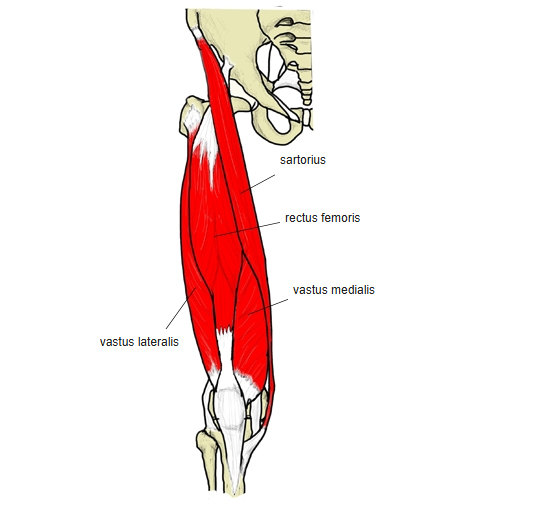
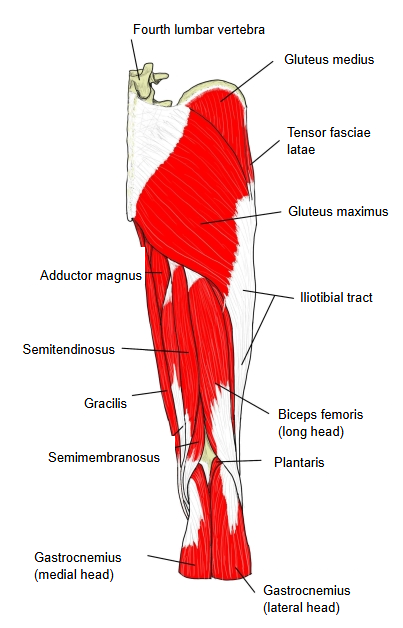
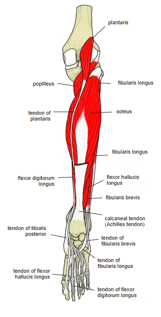
These are the main muscles that bend (flex) and straighten (extend) the knee joint.
| Muscle | Action at the knee | |
|---|---|---|
| Extensors | ||
| 1 | Rectus femoris | Extension |
| 2 | Vastus intermedius | Extension |
| 3 | Vastus medialis | Extension |
| 4 | Vastus lateralis | Extension |
| Flexors | ||
| 1 | Biceps femoris | Flexion, lateral rotation |
| 2 | Semimembranosus | Flexion, medial rotation |
| 3 | Semitendinosus | Flexion, medial rotation |
| 4 | Gastrocnemius | Flexion |
| 5 | Popliteus | Flexion, medial rotation |
| 6 | Gracilis | Flexion, medial rotation |
| 7 | Sartorius | Flexion, medial rotation |
| 8 | Plantaris | Flexion (weak) |
Note: Wikipedia groups these muscles by the compartments of the thigh, so its lists differ a little:
- It puts the Articularis genus with the extensors.
- It puts the Sartorius in the anterior (extensor) compartment.
- It puts the Gracilis in the medial compartment, neither with the extensors nor with the flexors.
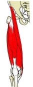 Right thigh (anterior view)
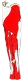 Right thigh (posterior view)
 Right leg (posterior view)
Ligaments
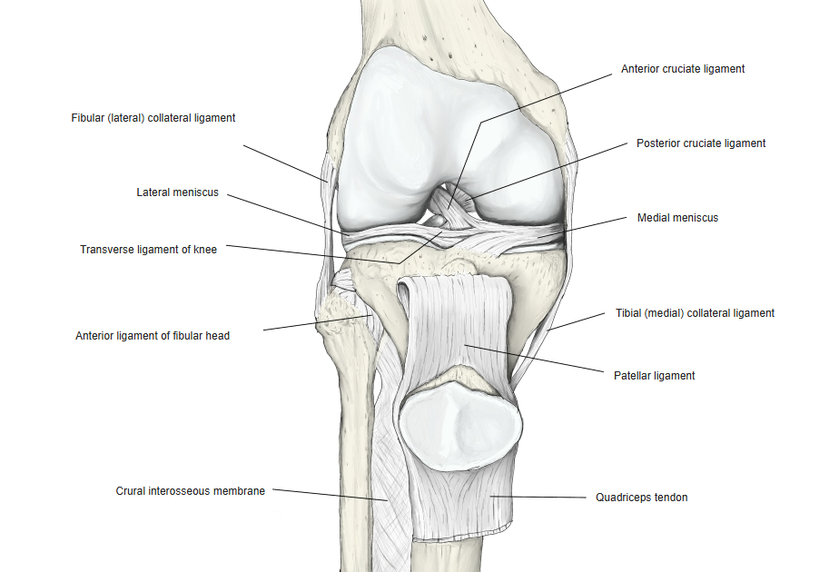
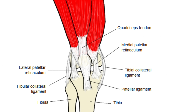
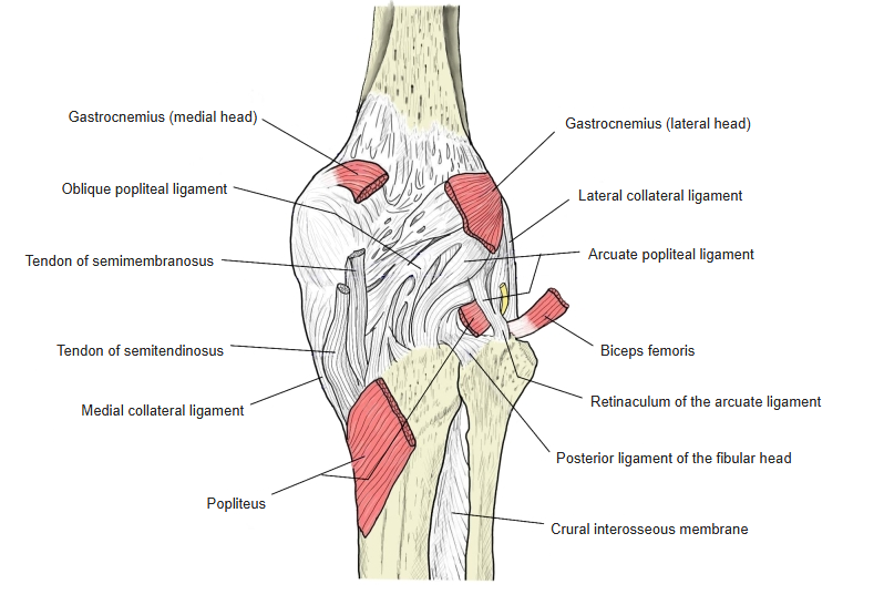
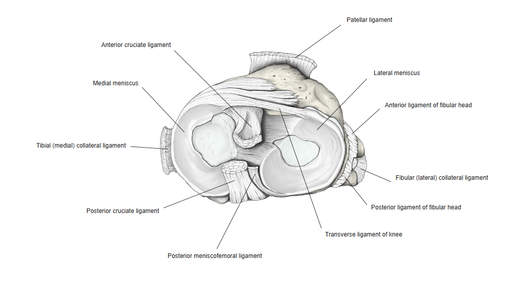
These ligaments strengthen the knee joint.