Joint capsule (Latin: capsula articularis)
Also called Articular capsule
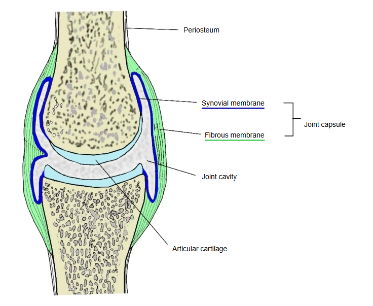
The joint capsule is the connective tissue that encloses a whole synovial joint; it is made of the two layers below. The space inside the capsule is the joint cavity, filled with clear synovial fluid.
capsule
- Fibrous membrane : (fibrous capsule) the outer layer; dense connective tissue, mainly collagen fibres with some elastic fibres.
- Synovial membrane : the inner layer; rich in blood vessels; produces the synovial fluid.
The fibrous membrane is attached all round the margins of the articular surfaces of the bones and is continuous with their periosteum, so it surrounds the joint completely. It gives the joint strength and is often thickened into capsular ligaments.
Inside the capsule, articular cartilage covers the ends of the bones. The fibrous membrane has a rich nerve supply, usually from branches of the nerves to the muscles acting on the joint.
Reference websites
[Wikipedia]
· Joint capsule, Wikipedia.
· Synovial joint, Wikipedia.
[Websites with illustrations]
· Joint capsule of a synovial joint (Gray's Anatomy) (Wikimedia Commons)

Related terms
- Synovial membrane
- Synovial fold
- Hip joint
- Knee joint
- Glenohumeral joint
- Zona orbicularis
- Temporomandibular joint
References and sources
- Further reading: Joint capsule, Wikipedia.
- Further reading: Synovial joint, Wikipedia.
- Federative International Programme for Anatomical Terminology (FIPAT). Terminologia Anatomica, 2nd ed. (TA2), 2019.