Olfactory bulb (Latin: bulbus olfactorius)
Also called Main olfactory bulb
· When it needs to be told apart from the accessory olfactory bulb, it is also called the main olfactory bulb.
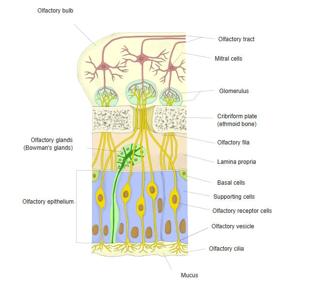
The olfactory nerve fibres, the axons of the olfactory cells in the olfactory epithelium of the nasal mucosa, pass up through the holes of the cribriform plate as the fila olfactoria and end in the bulb. The axons of the mitral and tufted cells leave it backwards as the olfactory tract and run to the olfactory cortex and the amygdala.
Its layers, from the surface inwards, are the olfactory nerve layer, glomerular layer, external plexiform layer, mitral cell layer, internal plexiform layer and granule cell layer.
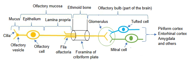
Note: these figures are based on "MR Imaging of Olfactory Bulbs and Tracts" (AJNR).
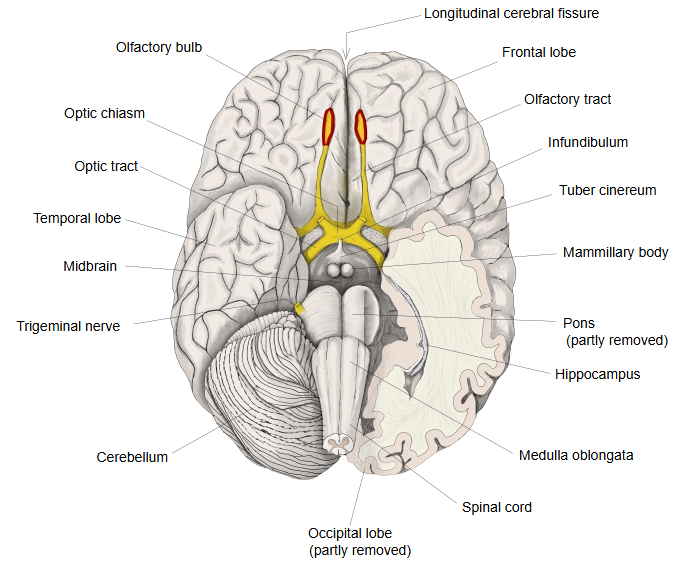
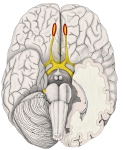 Brain (inferior view)
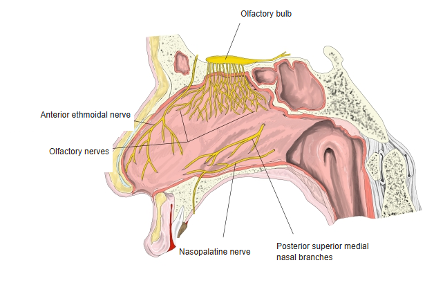
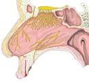 Olfactory bulb and nasal septum
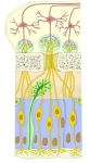 Olfactory region (section)
Reference websites
[Wikipedia]
· Olfactory bulb, Wikipedia.
[Websites with illustrations]
· Plan of olfactory neurons (Gray's Anatomy figure) (Wikimedia Commons)

· Olfactory bulb and olfactory epithelium (diagram) (Mammoth Memory)

· Olfactory system inside the head, with the olfactory bulb (Shutterstock)

Related terms
- Olfactory nerve
- Olfactory epithelium
- Olfactory receptor cells
- Olfactory organ
- Cribriform plate
- Nasal mucosa
- Brain
References and sources
- Further reading: Olfactory bulb, Wikipedia.
- Further reading: Olfactory system, Wikipedia.
- Federative International Programme for Anatomical Terminology (FIPAT). Terminologia Anatomica, 2nd ed. (TA2), 2019.