Posterior macula flava (Latin: macula flava posterior)
Anatomy textbooks describe them as follows.
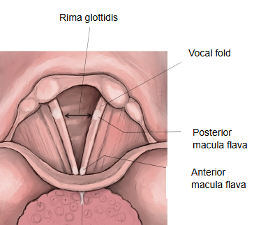
In the living larynx the vocal folds look whitish, but each end shows a slightly yellow spot (macula flava, "yellow spot"). The anterior macula flava lies at the front end, near the anterior commissure, and contains a small elastic sesamoid cartilage; the posterior macula flava marks the vocal process of the arytenoid cartilage. Both are dense masses of fibroblasts and elastic fibres at the two ends of the vocal ligament, and are thought to help the vocal fold withstand vibration.
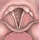 Anterior and posterior maculae flavae
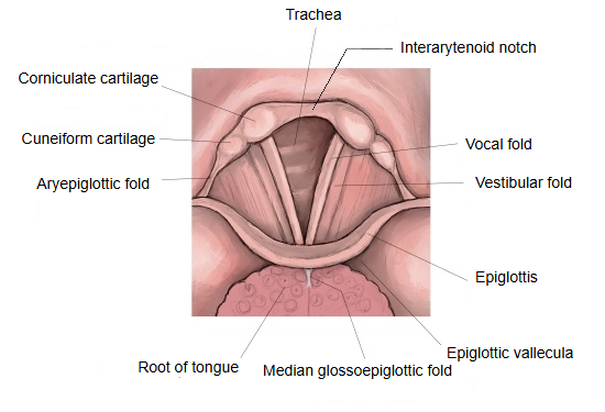
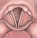 Larynx from above
Reference websites
[Wikipedia]
· Vocal folds, Wikipedia.
[Websites with illustrations]
· Structure of the vocal fold (Springer Nature)

· Coronal section of the larynx (Gray's Anatomy) (Wikimedia Commons)

Related terms
References and sources
- Further reading: Vocal folds, Wikipedia.
- Federative International Programme for Anatomical Terminology (FIPAT). Terminologia Anatomica, 2nd ed. (TA2), 2019.