Vocal fold (Latin: plica vocalis)
Also called Vocal cord or the true vocal cord or the true vocal fold
· As English Wikipedia also shows, the vocal fold and the vocal cord can be taken to be the same thing.
"In humans, the vocal cords, also known as vocal folds, are folds of throat tissues ..."
In other words, the vocal fold is one of the parts of the glottis.
· Each vocal fold runs from the inner angle of the thyroid cartilage in front to the vocal process of the arytenoid cartilage behind. The two folds and the gap between them (rima glottidis) together form the glottis; the laryngeal ventricle lies between each vocal fold and the vestibular fold above it, and the conus elasticus continues downward from the fold to the cricoid cartilage (cricothyroid ligament). The folds part during breathing and come together during speech; air pushed between them sets them vibrating, and their length and tension set the pitch of the voice.
· Other sources usually name two components of the vocal fold, the vocal ligament and the vocalis muscle, but
the conus elasticus, whose thickened upper edge is the vocal ligament, can also be counted as one of its components. (The oblique arytenoid muscle, sometimes suggested, lies on the back of the arytenoid cartilages and is not part of the fold.)
and
recesses
of the
larynx
- In the cavity
- Vestibular fold : a fold between the thyroid and arytenoid cartilages built on the vestibular ligament and the quadrangular membrane; also called the ventricular fold or false vocal cord.
- Vocal fold : a fold between the thyroid and arytenoid cartilages built on the vocal ligament and the conus elasticus; it produces the voice.
- At the inlet
- Aryepiglottic fold : stretched between the epiglottis and the arytenoid cartilage; it forms the two sides of the laryngeal inlet and carries two tubercles (cuneiform and corniculate).
- In front
- Median glosso-epiglottic fold : between the midline of the root of the tongue and the epiglottic cartilage; the only unpaired fold.
- Lateral glosso-epiglottic fold : on the front of the epiglottis, on the outer side of the epiglottic vallecula.
- Others
- Fold of the superior laryngeal nerve : in the wall of the piriform recess, where the course of the superior laryngeal nerve (from upper lateral to lower medial) raises a fold.
- Pharyngo-epiglottic fold : runs from the side wall of the pharynx to the outer side of the epiglottic cartilage; it lies in the laryngeal part of the pharynx.
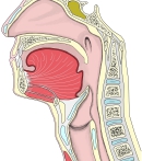 Nasal cavity to larynx (median section)
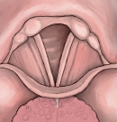 Larynx from above
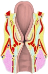 Larynx (coronal section)
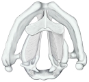 Laryngeal cartilages from above
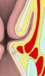 Vestibular and vocal folds (coronal section)
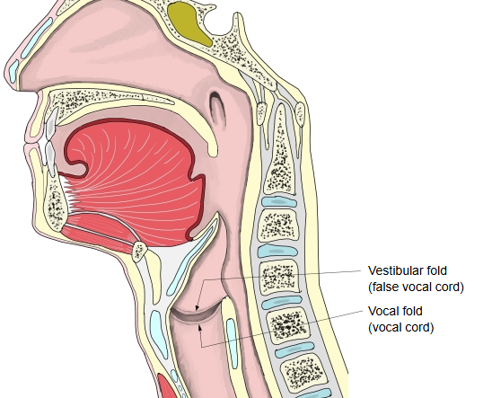
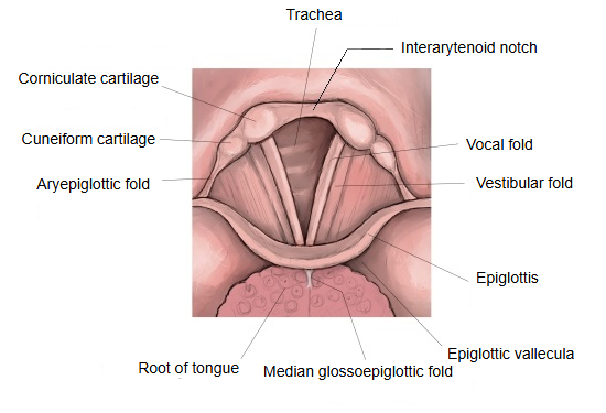
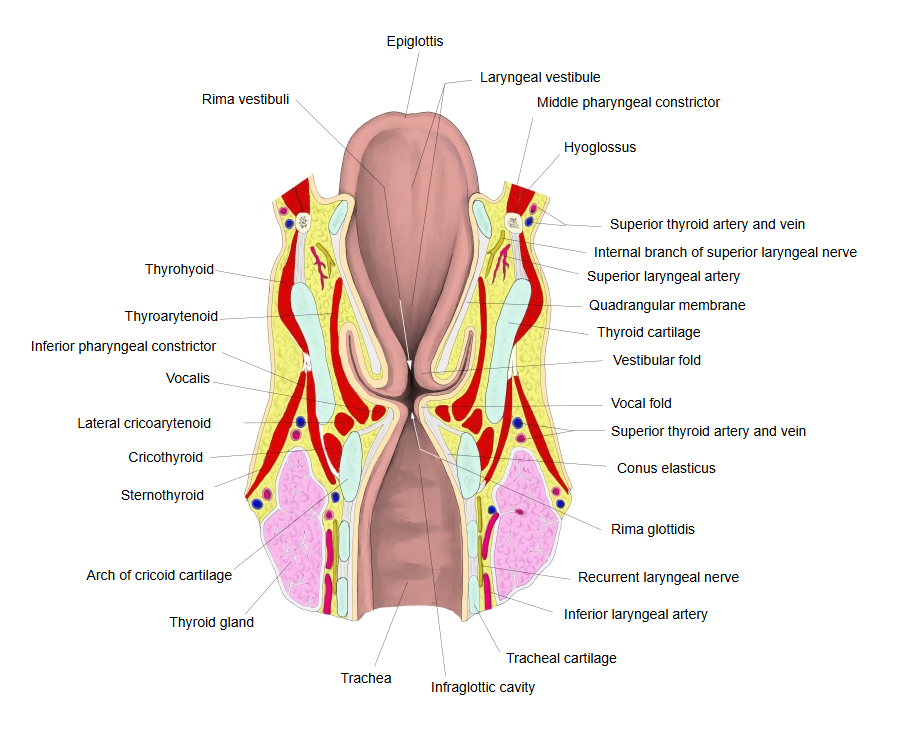
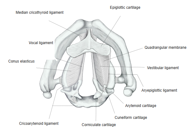
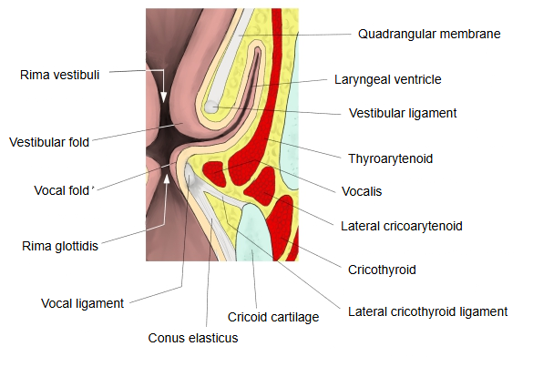
A standard anatomy textbook adds the following.
In the living body the vocal folds look fairly white (pearly white: the free edge is covered by stratified squamous epithelium and has few vessels). Their front end and their cartilaginous back part look slightly yellowish, because of the sesamoid cartilage and tissue rich in elastic fibres in front and the vocal process of the arytenoid cartilage behind (the anterior and posterior maculae flavae).
Reference websites
[Wikipedia]
· Vocal cords, Wikipedia.
[Websites with illustrations]
· Vocal folds open, seen from above (squarespace-cdn.com)
· The larynx in coronal section (vestibular and vocal folds labelled) (ohsu.edu)

· Coronal section of the larynx: vestibular and vocal ligaments, conus elasticus (Springer Nature)

· The larynx: midsagittal and endoscopic views (vocalprocess.co.uk)

Where it is found
Related terms
- Vocal ligament
- Glottis
- Rima glottidis
- Vestibular fold
- Laryngeal ventricle
- Cricothyroid ligament
- Larynx
References and sources
- Further reading: Vocal cords, Wikipedia.
- Federative International Programme for Anatomical Terminology (FIPAT). Terminologia Anatomica, 2nd ed. (TA2), 2019.