Thyroid articular surface (Latin: Facies articularis thyroidea)
Also called the articular facet for thyroid cartilage
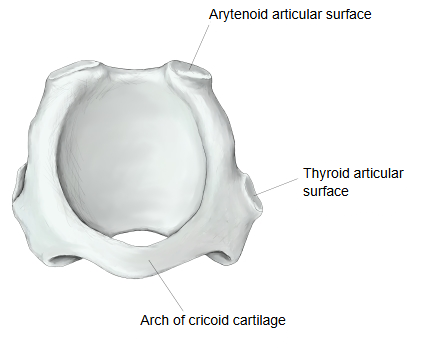
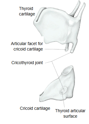
· It is a small, round joint facet, one on each side, on the outer surface of the cricoid cartilage where the lamina meets the arch. It corresponds to the articular facet on the tip of the inferior horn of the thyroid cartilage.
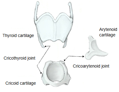
Together the two facets form the cricothyroid joint, a synovial joint. At this joint the thyroid cartilage tilts forwards and back on the cricoid cartilage, which changes the tension of the vocal folds (see cricothyroid). The other paired facet of the cricoid cartilage, the arytenoid articular surface, lies on the upper border of the lamina.
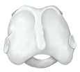 Cricoid cartilage (posterior view)
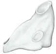 Cricoid cartilage (left lateral view)
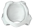 Cricoid cartilage (superior view)
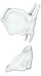 Cricothyroid joint
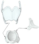 Joints of the larynx
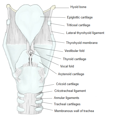
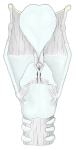 Larynx (posterior view)
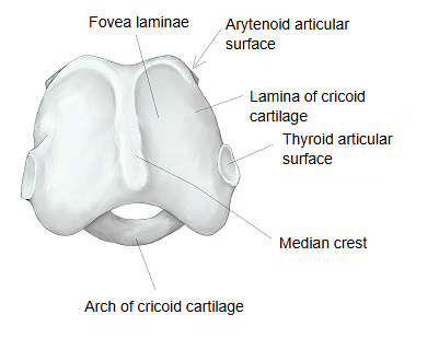
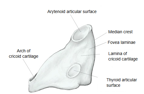
The cricoid cartilage as a whole:
It is an unpaired piece of hyaline cartilage below the thyroid cartilage, joined below to the first tracheal cartilage by the cricotracheal ligament. It is shaped like a signet ring, rising from front to back: the narrow front part is the arch, and the back part, widened into a square plate, is the lamina. The back of the lamina carries the median crest and, on each side of it, the fovea laminae, where the posterior cricoarytenoid muscle arises. The small, round thyroid articular surfaces lie where the lamina meets the arch, and the oval arytenoid articular surfaces lie on each side of the upper border of the lamina, where the arytenoid cartilages sit.
Related terms
- Arytenoid articular surface
- Fovea laminae
- Inferior horn of thyroid cartilage
- Arytenoid cartilage
- Larynx
- Cricothyroid ligament
- Cricothyroid
References and sources
- Further reading: Cricoid cartilage, Wikipedia.
- Federative International Programme for Anatomical Terminology (FIPAT). Terminologia Anatomica, 2nd ed. (TA2), 2019.