Latin: musculus iliacus
Overview
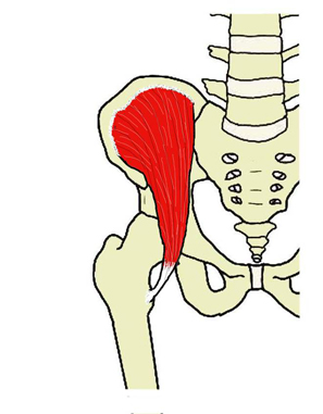
- A flat, triangular muscle that fills the iliac fossa; it is covered by the iliac fascia.
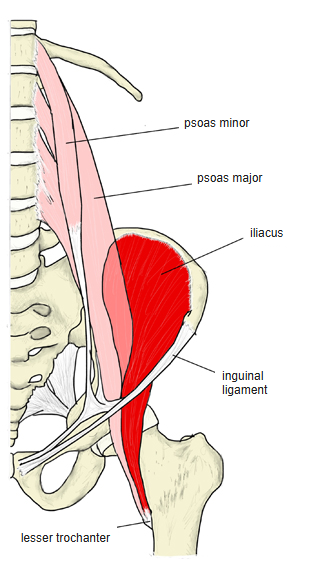
- With the psoas major it forms the iliopsoas, which passes under the inguinal ligament through the muscular lacuna.
- Acts only on the hip joint, whereas the psoas major also crosses the joints of the lumbar spine.
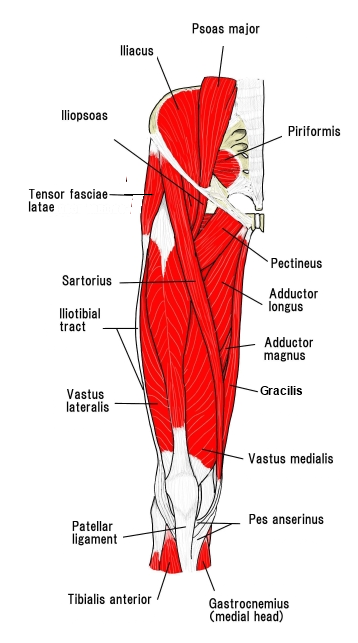
- Continuous in places with the psoas major, rectus femoris, sartorius, vastus medialis, pectineus and tensor fasciae latae.
Position in the classification
Lower Limb › Hip › Inner hip muscles (iliopsoas)
- Iliacus
- Psoas major
- Psoas minor
Illustrations
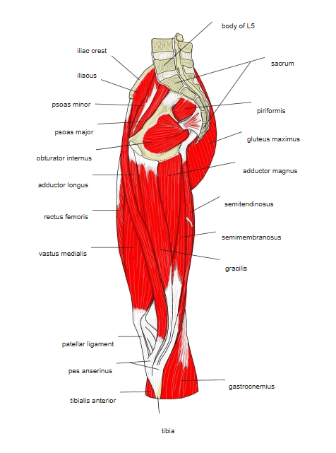
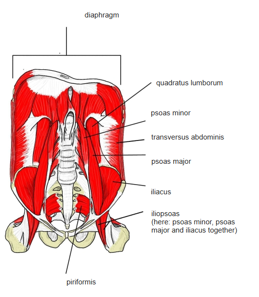
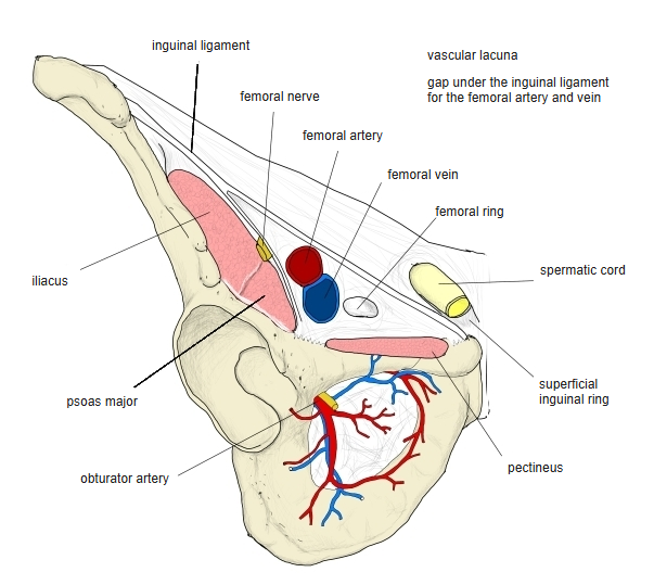
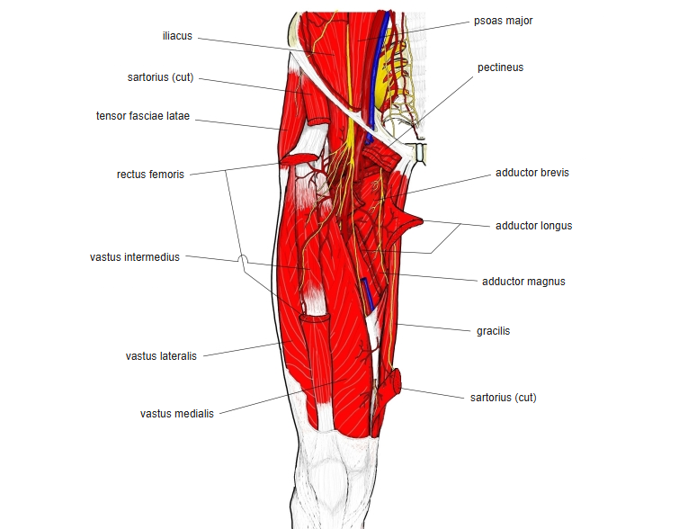
Origin / insertion
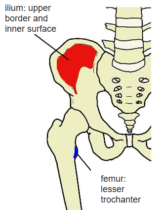
- Origin: upper two-thirds of the iliac fossa, inner lip of the iliac crest, ala of the sacrum, and the anterior sacroiliac and iliolumbar ligaments; in front it reaches the anterior superior and anterior inferior iliac spines. The fibres run down and medially
- Insertion: lesser trochanter of the femur, through the lateral side of the psoas major tendon; some fibres continue onto the femur just below and in front of the lesser trochanter (some sources add the hip joint capsule)
Action
- Flexes the hip; with the femur fixed, helps the psoas major to tilt the pelvis forward
- Laterally rotates the hip
Innervation
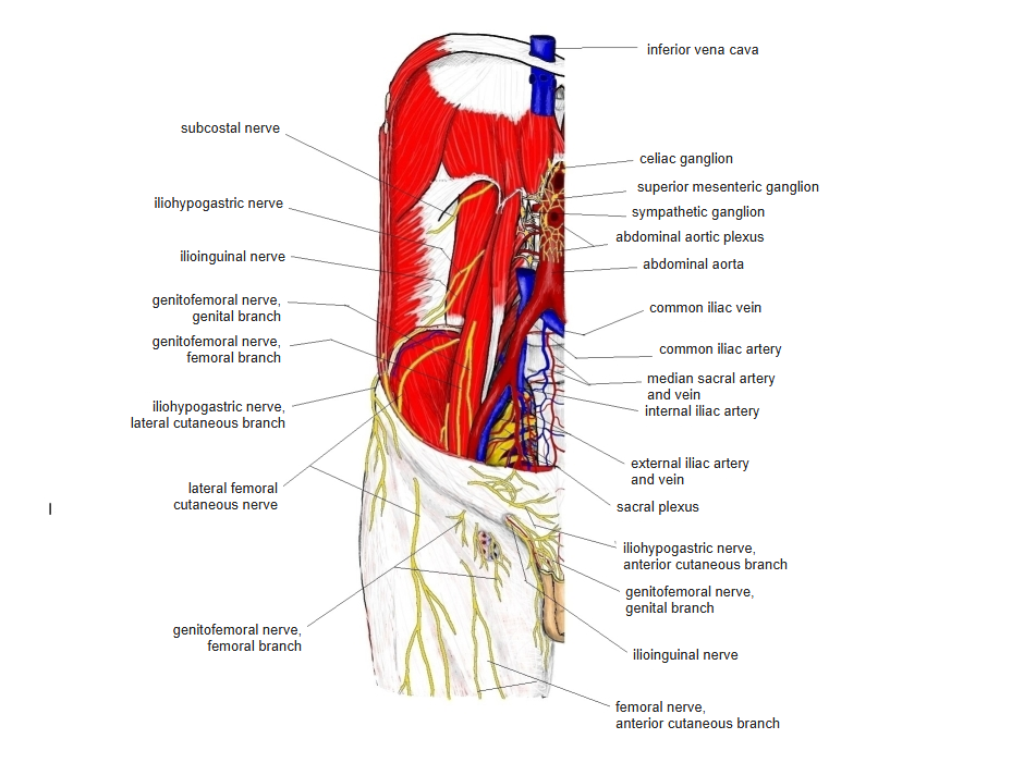
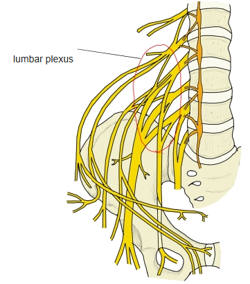
- Femoral nerve (L2–L3; some sources L2–L4), often also by direct branches of the lumbar plexus
Blood supply
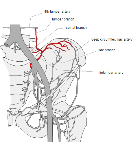
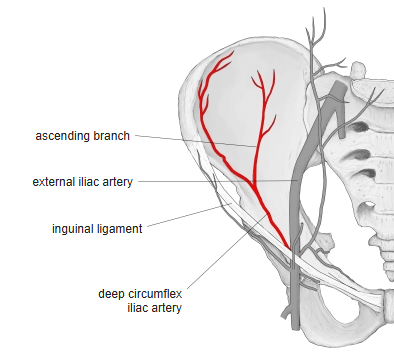
- Iliolumbar artery, deep circumflex iliac artery
Related terms
- Psoas major
- Psoas minor
- Iliopsoas
- Iliac fossa
- Lesser trochanter
- Lumbar plexus
- Femoral nerve
References and sources
- Further reading: Iliacus muscle, Wikipedia.