Suboccipital triangle (Latin: trigonum suboccipitale)
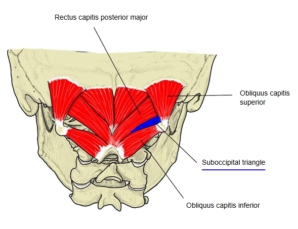
A small triangular space deep in the back of the upper neck (the occipital region), bounded by three of the suboccipital muscles: rectus capitis posterior major above and medially, obliquus capitis superior above and laterally, and obliquus capitis inferior below and laterally. It contains the third part of the vertebral artery, lying in the groove on the posterior arch of the atlas, and the suboccipital nerve (dorsal ramus of the first cervical nerve). Its floor is the posterior atlanto-occipital membrane and the posterior arch of the atlas; its roof is dense fibrofatty tissue under semispinalis capitis. The greater occipital nerve appears below obliquus capitis inferior and crosses the roof of the triangle on its way up to the scalp; it is not inside the triangle itself.
Related terms
- Rectus capitis posterior major
- Obliquus capitis superior
- Obliquus capitis inferior
- Vertebral artery
- Suboccipital nerve
- Greater occipital nerve
- Posterior atlanto-occipital membrane
References and sources
- Further reading: Suboccipital triangle, Wikipedia.
- Federative International Programme for Anatomical Terminology (FIPAT). Terminologia Anatomica, 2nd ed. (TA2), 2019.