Choroid glomus (Latin: glomus choroideum)
Also called Choroid enlargement, choroid skein
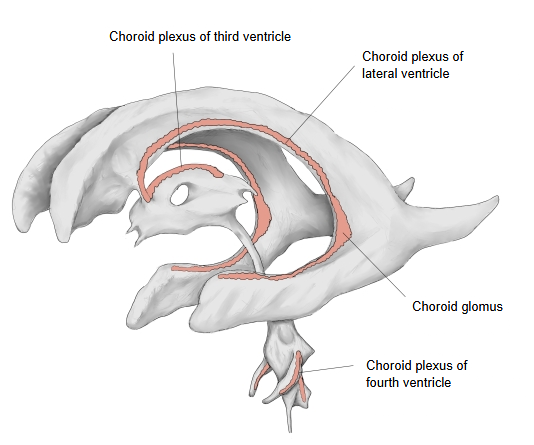
The choroid plexus of the lateral ventricle is attached by its choroid line (taenia) along the stria terminalis. It begins at the front end of the inferior horn of the lateral ventricle, curves round through the central part and reaches the interventricular foramen, where the plexuses of both sides meet, turn backwards and continue into the choroid plexus of the third ventricle. The part where it passes from the inferior horn into the central part is especially thick; this is the choroid glomus (choroid skein), the bulkiest part of the plexus.
The choroid glomus is the enlarged part of the choroid plexus in the atrium of the lateral ventricle, where the body, the posterior horn and the inferior horn meet. It often becomes partly calcified with age and then appears white on computed tomography (CT).
Like the rest of the choroid plexus, the choroid glomus produces cerebrospinal fluid. Its calcification usually shows as bright spots on both sides and is a normal finding. It is supplied by the anterior choroidal artery and the posterior choroidal arteries, and drained by the superior choroid vein.
Related terms
- Choroid plexus
- Inferior horn of lateral ventricle
- Posterior horn of lateral ventricle
- Third ventricle
- Anterior choroidal artery
- Superior choroid vein
References and sources
- Further reading: Choroid plexus, Wikipedia.
- Federative International Programme for Anatomical Terminology (FIPAT). Terminologia Anatomica, 2nd ed. (TA2), 2019.