Medial patellar retinaculum (Latin: retinaculum patellae mediale)
Also called: tibial patellar retinaculum
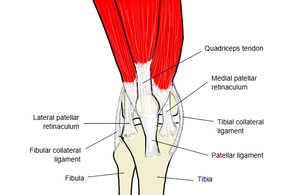
It is one of the structures that stabilise the knee joint.
It is a strong, tendon-like fibrous sheet on the front of the knee, medial to the patella and the patellar ligament. It arises from the medial side of the tendon of quadriceps femoris and the medial border of the patella, and runs down to the front of the medial condyle of the tibia.
On both sides of the patella and the patellar ligament, the aponeuroses of vastus medialis and vastus lateralis join the deep fascia of the thigh that covers them, forming strong, sheet-like bundles of longitudinal fibres that reinforce the joint capsule. These are the medial patellar retinaculum and the lateral patellar retinaculum. The medial retinaculum strengthens the anteromedial side of the capsule, in front of the tibial collateral ligament. With the lateral retinaculum on the other side of the patellar ligament, it helps hold the patella in its groove on the femur and resists lateral displacement of the patella.
Reference websites
[Wikipedia]
· Knee, Wikipedia.
[Websites with illustrations]
· Knee joint, anterior view, with the patellar retinacula (slide 12) (SlideShare)

Related terms
- Knee joint
- Patella
- Patellar ligament
- Vastus medialis
- Quadriceps femoris
- Tibial collateral ligament
- Lateral patellar retinaculum
References and sources
- Further reading: Knee, Wikipedia.
- Federative International Programme for Anatomical Terminology (FIPAT). Terminologia Anatomica, 2nd ed. (TA2), 2019.