Latin: musculus vastus lateralis
Overview
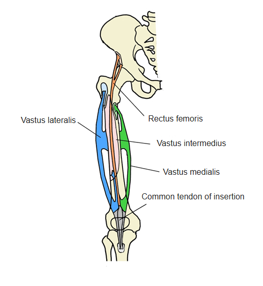
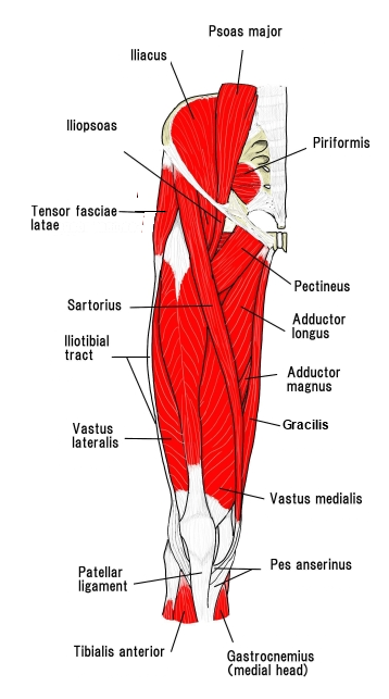
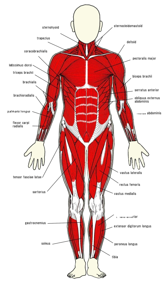
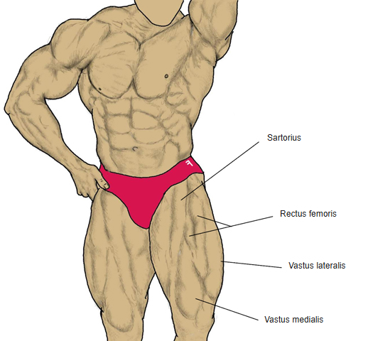
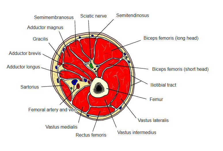
- The largest of the four heads of the quadriceps femoris, on the lateral side of the thigh.
- Its belly ends about two finger-breadths above the patella; the vastus medialis reaches lower.
- Fibres at its back running obliquely downward and forward are often called the oblique head.
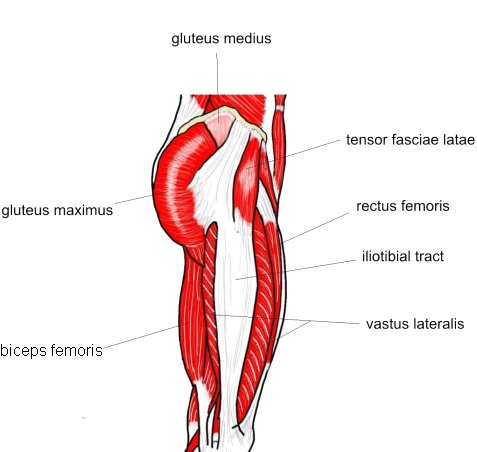
- Joined to many neighbouring muscles, e.g. the biceps femoris (short head), gluteus maximus, rectus femoris and vastus intermedius.
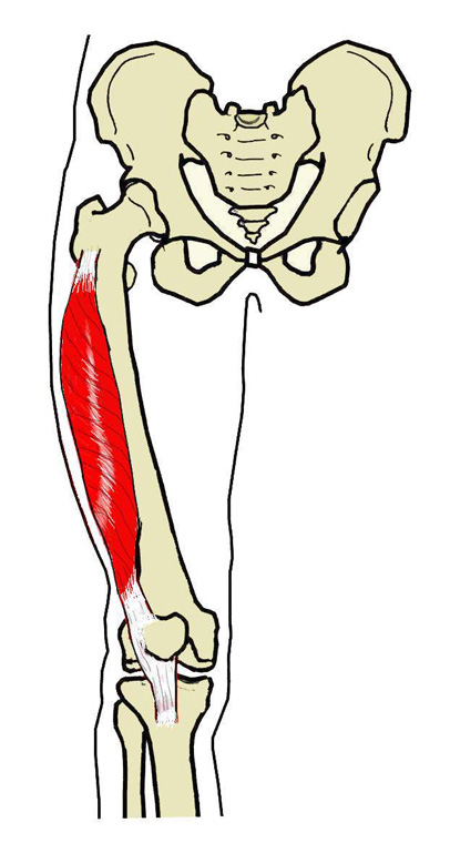
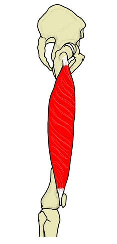
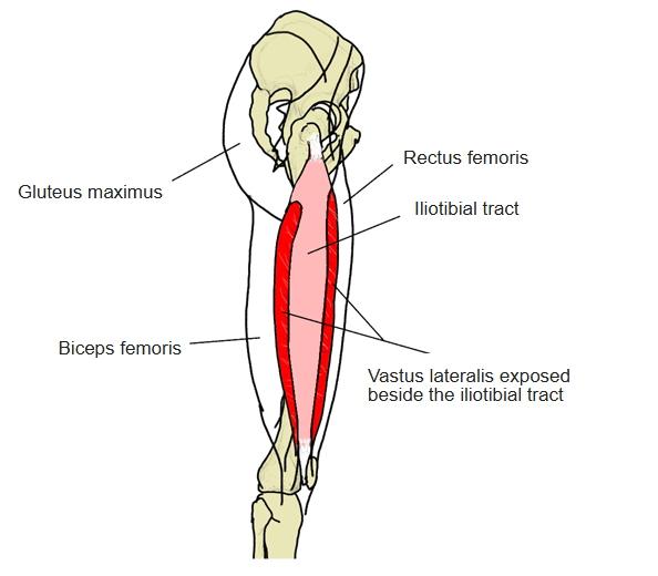
Position in the classification
Lower Limb › Thigh › Extensor group › Quadriceps femoris
- Rectus femoris
- Vastus medialis
- Vastus lateralis
- Vastus intermedius
Illustrations
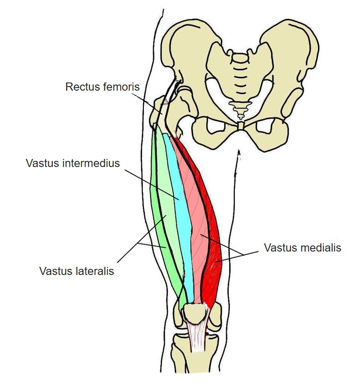
Origin / insertion
- Origin: lateral surface of the greater trochanter, lateral lip of the linea aspera (femur)
- Insertion: superior border and lateral side of the patella (common quadriceps tendon), and via the patellar ligament to the tibial tuberosity; some tendon fibres spread into the patellar retinaculum
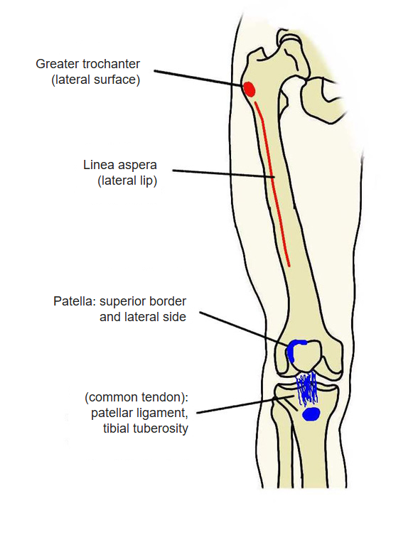
Many sources give the patella as the insertion; the patellar ligament and tibial tuberosity are included here as the common tendon of the quadriceps.
Action
- Extends the knee (with the other heads of the quadriceps)
Innervation
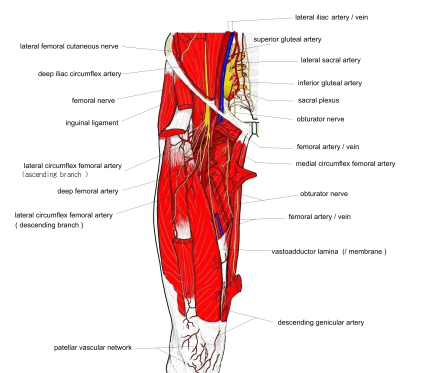
- Femoral nerve (L2–L4)
Blood supply
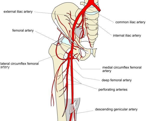
- Lateral circumflex femoral artery
Related terms
- Rectus femoris
- Vastus medialis
- Vastus intermedius
- Iliotibial tract
- Femoral nerve
- Lateral circumflex femoral artery
References and sources
- Further reading: Vastus lateralis muscle, Wikipedia.