Anterior horn of lateral ventricle (Latin: cornu anterius ventriculi lateralis)
Also called the frontal horn
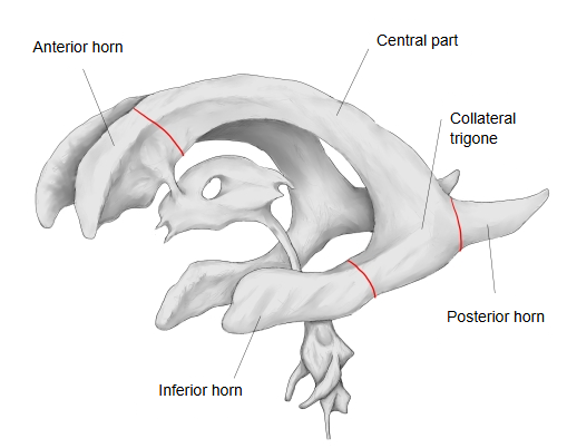
The anterior horn (of the lateral ventricle) is
The main parts of the lateral ventricle are as follows.
- Anterior horn : lies in the frontal lobe.
- Central part : lies in the parietal lobe.
- Inferior horn : lies in the temporal lobe; its tip almost reaches the front end of the temporal lobe. The longest horn.
- Posterior horn : lies in the occipital lobe.
Walls
The walls of the anterior horn are formed as follows.
· Medial wall: the septum pellucidum (its front part, which separates the two anterior horns).
· Upper wall (roof and front): the genu of the corpus callosum and part of the trunk of the corpus callosum.
· Inferolateral wall: the head of the caudate nucleus, which bulges into the horn from the side.
· Floor: the rostrum of the corpus callosum. Sources differ in how they name the walls: some call the caudate side the lateral wall and the rostrum the floor, others join them as the inferolateral wall.
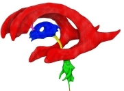 Ventricles: left side (coloured)
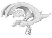 Ventricles: left side
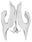 Ventricles: from above
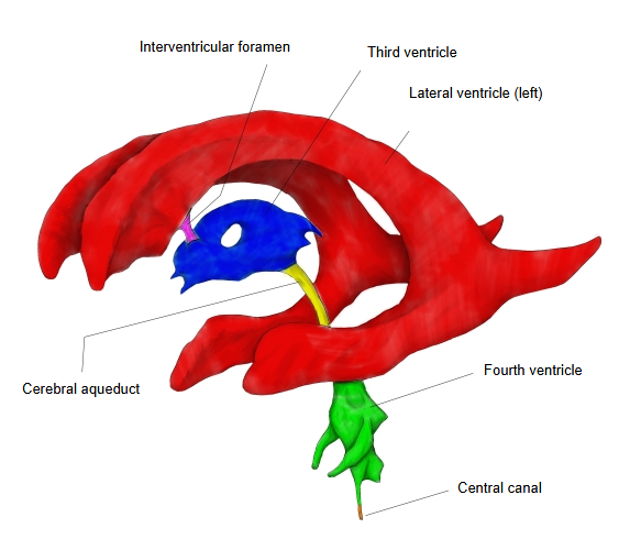
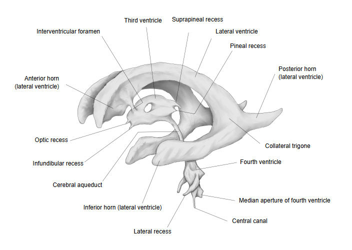
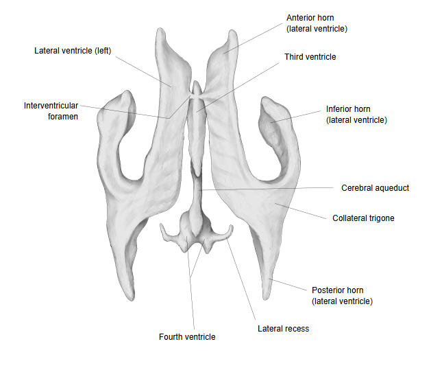
The anterior horn passes forwards, outwards and slightly downwards from the interventricular foramen, through which it opens into the third ventricle, into the frontal lobe, and curves round the front of the caudate nucleus. Its floor is formed by the upper surface of the rostrum of the corpus callosum; it is bounded medially by the front part of the septum pellucidum and laterally by the head of the caudate nucleus, and its tip reaches the back of the genu of the corpus callosum. In a coronal section it is roughly triangular. Unlike the central part and the inferior horn, it contains no choroid plexus.
Reference websites
[Wikipedia]
· Lateral ventricles, Wikipedia.
· Ventricular system, Wikipedia.
Related terms
- Cerebral ventricles
- Posterior horn of lateral ventricle
- Inferior horn of lateral ventricle
- Third ventricle
- Choroid plexus
- Anterior vein of septum pellucidum
- Brain
References and sources
- Further reading: Lateral ventricles, Wikipedia.
- Further reading: Ventricular system, Wikipedia.
- Federative International Programme for Anatomical Terminology (FIPAT). Terminologia Anatomica, 2nd ed. (TA2), 2019.