Patellar anastomosis (Latin: rete patellare)
Also called the patellar network or the patellar arterial network
The patellar anastomosis is the part of the genicular anastomosis that lies in front of the patella.
Genicular anastomosis: the arterial network that surrounds the knee joint capsule in front and behind, formed by the superior medial, superior lateral, inferior medial and inferior lateral genicular arteries, the descending genicular artery, the anterior tibial recurrent artery and other arteries
· It lies on and around the anterior surface of the patella, in front of the knee joint. Many small vessels from this network enter the patella through its anterior surface and supply most of the bone.
· Sources differ: some books and websites use "patellar network" or "patellar anastomosis" for the whole genicular anastomosis, not only its part on the patella.
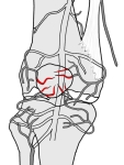 Patellar anastomosis
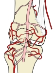 Arteries around the knee (anterior view)
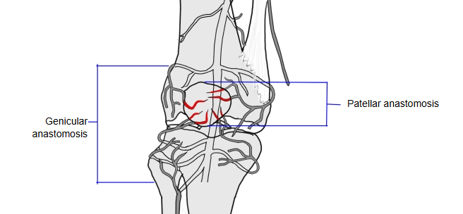
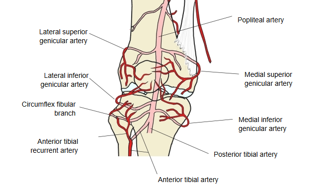
Reference websites
[Wikipedia]
· Patellar network, Wikipedia.
[Websites with illustrations]
· Arteries around the patella (anterior view) (Kenhub)

· Arteries of the knee with the patella (labelled) (Fine Art America)

Related terms
- Infrapatellar synovial fold
- Infrapatellar fat pad
- Genicular anastomosis
- Patellar region
- Popliteal region
- Adipose folds
- Patella
- Medial superior genicular artery
- Lateral superior genicular artery
- Medial inferior genicular artery
- Lateral inferior genicular artery
- Anterior tibial recurrent artery
References and sources
- Further reading: Patellar network, Wikipedia.
- Federative International Programme for Anatomical Terminology (FIPAT). Terminologia Anatomica, 2nd ed. (TA2), 2019.