Infrapatellar synovial fold (Latin: plica synovialis infrapatellaris)
Also called the ligamentum mucosum
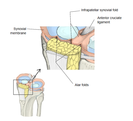
The infrapatellar synovial fold is a fold of the synovial layer of the articular capsule of the knee joint, and the largest synovial fold of the knee. The synovial layer is attached to the outer margins of the medial and lateral menisci (their articular surfaces are not covered by synovium) and covers the front and sides of the anterior and posterior cruciate ligaments; it also reaches into the joint cavity to form this fold. The fold arises from the front of the intercondylar fossa of the femur, runs in front of the anterior cruciate ligament and reaches the infrapatellar fat pad below the apex of the patella. At its lower end it spreads out on each side into the alar folds, which contain much fat (the infrapatellar fat pad) and reach the sides of the patella.
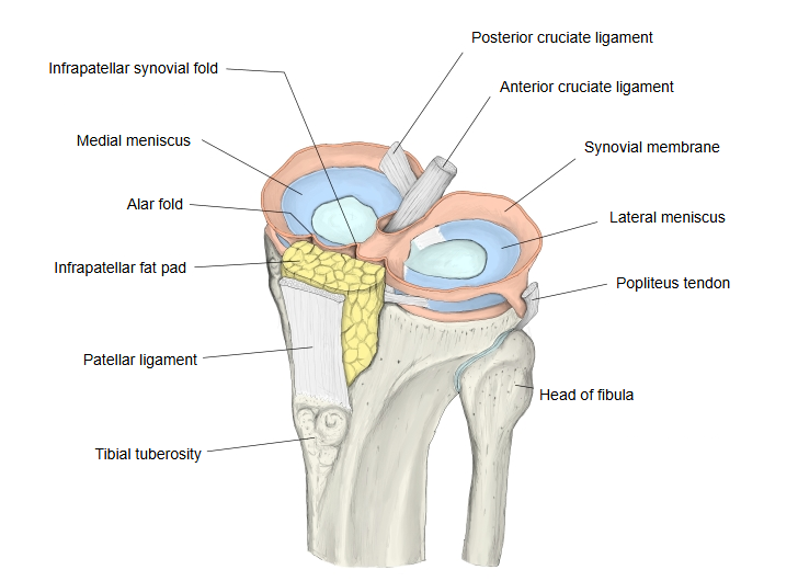
It is a remnant of a midline partition that, before birth, runs from the back wall of the capsule (containing the cruciate ligaments) to the front wall below the patella and divides the joint cavity into right and left halves. How much of it remains varies; its main remnant is the infrapatellar synovial fold. In adults there is often a gap between the fold and the cruciate ligament.
Its size varies: it may be a broad band or only a single thin cord of synovial membrane crossing the joint cavity from the fat pad to the anterior cruciate ligament. It is seen during arthroscopy of the knee and is usually of no clinical importance.
Related terms
- Infrapatellar fat pad
- Patellar anastomosis
- Genicular anastomosis
- Patellar region
- Popliteal region
- Adipose folds
- Synovial fold
- Synovial membrane
- Knee joint
- Intercondylar fossa
- Anterior cruciate ligament
- Articular cavity
References and sources
- Further reading: Plica syndrome, Wikipedia.
- Federative International Programme for Anatomical Terminology (FIPAT). Terminologia Anatomica, 2nd ed. (TA2), 2019.