Pterygospinous foramen (Latin: foramen pterygospinosum)
Also called the foramen of Civinini or the the bony bar that forms it is the pterygospinous bar
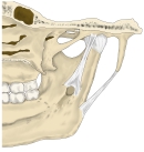 Pterygospinous bar and foramen
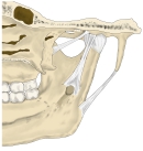 Ligaments near the temporomandibular joint
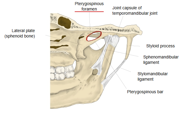
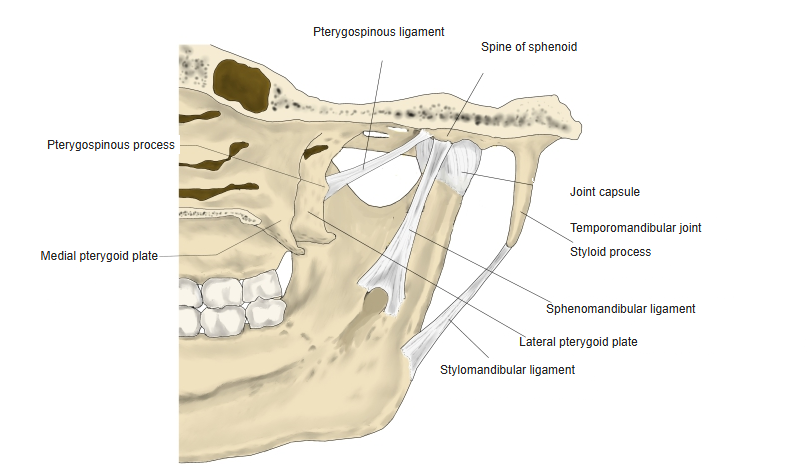
Small branches of the mandibular nerve to the muscles of mastication, such as the nerve to medial pterygoid, and small vessels may pass through it; sources differ on the details.
A bar or foramen is found in only a minority of skulls. It may get in the way of a needle aimed at the mandibular nerve near the foramen ovale and may press on nearby nerves. Another ligament in this area that can ossify is the sphenomandibular ligament.
Reference websites
[Wikipedia]
· Pterygospinous ligament, Wikipedia.
· Sphenoid bone, Wikipedia.
[Websites with illustrations]
· Base of the skull, outer surface (Gray's Anatomy) (Wikimedia Commons)

· Sphenoid bone, anterior and inferior surfaces (Gray's Anatomy) (Wikimedia Commons)

Related terms
- Pterygospinous ligament
- Pterygoid process
- Spine of sphenoid
- Foramen ovale
- Sphenoid bone
- Sphenomandibular ligament
References and sources
- Further reading: Pterygospinous ligament, Wikipedia.
- Federative International Programme for Anatomical Terminology (FIPAT). Terminologia Anatomica, 2nd ed. (TA2), 2019.