Foramen ovale (Latin: foramen ovale)
Also called Foramen ovale of sphenoid (not the foramen ovale of the fetal heart)
The foramen ovale is:
· The opening between the right and left atria of the fetal heart is also called the foramen ovale; it is a different structure, which closes after birth and leaves a trace, the oval fossa.
Nerves, arteries and veins passing through it
The nerves and vessels (vessels) that pass through the foramen ovale are listed below. Sources differ somewhat on what passes through it, and some textbooks mention only the mandibular nerve (third division of the trigeminal nerve).
| 1 | Nerves | · Mandibular nerve (third division of the trigeminal nerve, V3) · Motor root of the trigeminal nerve · Lesser petrosal nerve (from the glossopharyngeal nerve); sources differ, and it may instead pass through a small separate opening nearby |
| 2 | Artery | · Accessory meningeal artery (a branch of the maxillary artery or of the middle meningeal artery) |
| 3 | Veins | · An emissary vein, usually a small plexus (the venous plexus of the foramen ovale), linking the cavernous sinus with the pterygoid plexus · Middle meningeal veins (occasionally) |
Note: most textbooks send the middle meningeal veins with the middle meningeal artery through the foramen spinosum; only occasionally does a branch of the vein pass through the foramen ovale.
Because the mandibular nerve leaves the skull here just below the trigeminal ganglion, the foramen ovale is the usual route for needles in procedures on the ganglion for trigeminal neuralgia.
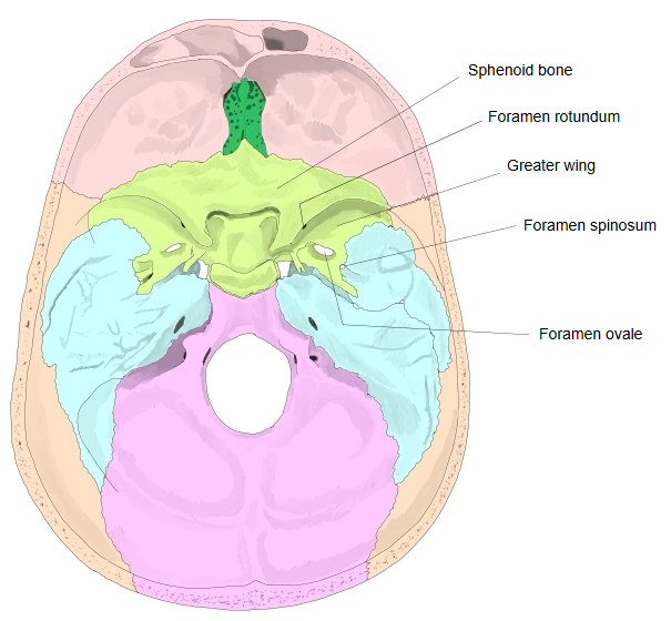
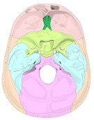 Cranial base (internal surface, bones coloured)
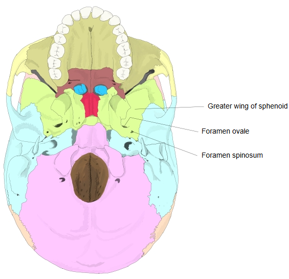
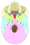 Cranial base (external surface)
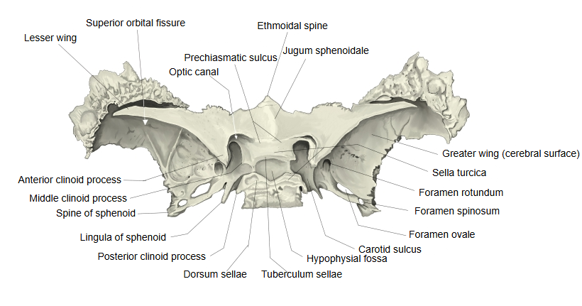
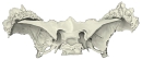 Sphenoid bone (superior)
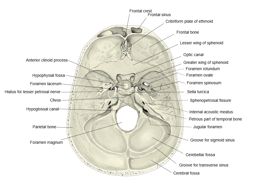
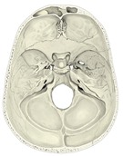 Cranial base (internal surface)
Reference websites
[Wikipedia]
· Foramen ovale (skull), Wikipedia.
· Mandibular nerve, Wikipedia.
[Websites with illustrations]
· Foramen ovale (Kenhub)
· Bony landmarks of the sphenoid wings and pterygoid process (TeachMeAnatomy)

· Skull, inferior view, with the foramen ovale (GetBodySmart)

Where it is found
Related terms
- Greater wing
- Foramen rotundum
- Superior orbital fissure
- Sella turcica
- Pterygoid process
- Sphenoid bone
References and sources
- Further reading: Foramen ovale (skull), Wikipedia.
- Further reading: Mandibular nerve, Wikipedia.
- Federative International Programme for Anatomical Terminology (FIPAT). Terminologia Anatomica, 2nd ed. (TA2), 2019.