Dentinal tubules (Latin: tubuli dentinales)
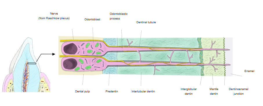
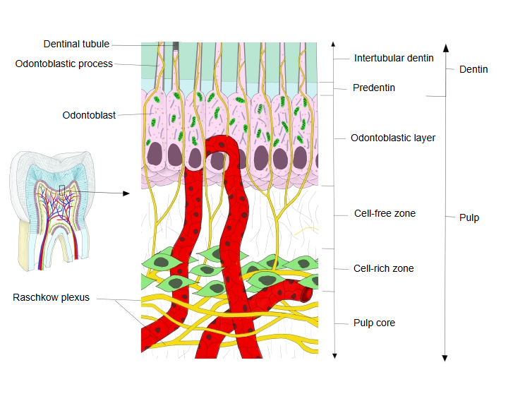
Note: illustrations of the course of the dentinal fiber show two patterns:
1. 1. It reaches the border with the enamel or the cementum
(that is, it runs the whole length of the tubule).
2. 2. It stops partway along the tubule. Example illustration 1, example illustration 2. How far the process reaches is still debated; sources differ.
· The tubules are walled by highly mineralized peritubular dentin; the dentin between them is intertubular dentin. Near the pulp they pass through the unmineralized predentin.
Note: some illustrations on the web show arteries and veins as well as nerves running in the dentinal tubules.
This is not correct: the tubules contain no blood vessels.
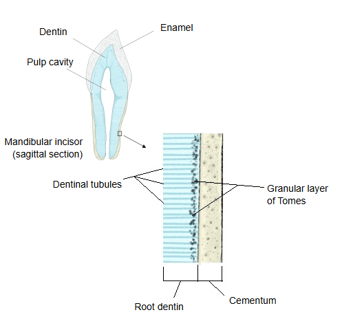
· In the crown the tubules follow a gentle S-shaped course, pass through the interglobular dentin and reach the dentinoenamel junction; in the root they are straighter and end at the granular layer of Tomes under the cementum.
· They take part in the sensitivity of the tooth: movement of the fluid in the tubules is thought to explain why exposed dentin feels cold water or sweet food (hydrodynamic theory).
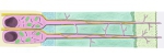 Structure of dentin
 Tubules in the root dentin
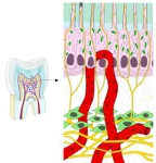 Odontoblasts and their processes


Reference websites
[Wikipedia]
· Dentin, Wikipedia.
[Websites with illustrations]
· Dentin (Pocket Dentistry)
· Gray's Anatomy, figure 1005: vertical section of a tooth (Wikimedia Commons)

· Tooth section showing enamel, dentin and dentinal tubules (Shutterstock)

Related terms
- Dentinal fiber
- Teeth
- Dental pulp
- Predentin
- Intertubular dentin
- Interglobular dentin
- Raschkow plexus
- Dentinoenamel junction
References and sources
- Further reading: Dentin, Wikipedia.
- Further reading: Odontoblast, Wikipedia.
- Federative International Programme for Anatomical Terminology (FIPAT). Terminologia Anatomica, 2nd ed. (TA2), 2019.