Platelet
Also called the thrombocyte
One of the cellular components of the blood: a small fragment of cell, without a nucleus, that helps to stop bleeding.
More details
Like the white blood cells and red blood cells, platelets are derived from the haematopoietic stem cells of the red bone marrow, but they have no nucleus. They are also called thrombocytes, and the abbreviation PLT is often used in blood tests. Their main features are as follows.
| 1 | Shape | Disc-shaped. At rest the surface of a platelet is smooth, but when bleeding occurs its cytoskeleton changes and it puts out many long projections, becoming spiky.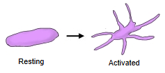 |
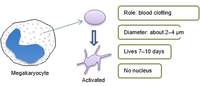
| 2 | Size | The smallest of the formed elements of the blood: about 2–3 µm across when inactive (sources give slightly different ranges). 1 µm = 1/1000 mm.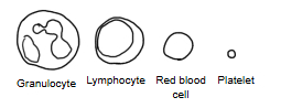 |
| 3 | Formation | Platelets are pieces of the cytoplasm of mature megakaryocytes, large cells of the bone marrow; each megakaryocyte breaks up into thousands of platelets. Haematopoietic stem cell → myeloid stem cell → megakaryoblast → megakaryocyte → platelets 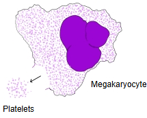 |
| 4 | Number | About 150,000–400,000 per µL (per mm³) of blood |
| 5 | Lifespan | About 8–9 days on average. Old platelets are removed mainly in the spleen, and also in the liver. |
Function
Blood clotting and haemostasis
The function of platelets is haemostasis, stopping bleeding. The process by which platelets stop bleeding is, in simplified form, as follows.
| 1 | The vessel wall is damaged and the collagen of the tissue beneath the endothelial cells is exposed. | 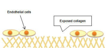 |
| 2 | The clotting factor von Willebrand factor (vWF) in the blood binds to the exposed collagen. At the same time the clotting (coagulation) cascade is set going at the injured site. | 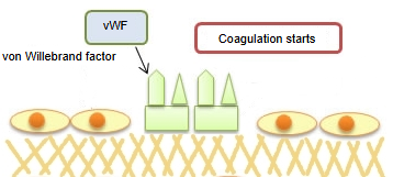 |
| 3 | Platelets bind to the vWF through the platelet membrane glycoprotein receptor GPIb (the GPIb-IX-V complex) and stick to the collagen (platelet adhesion, the first step of primary haemostasis, the forming of the platelet plug). | 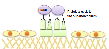 |
| 4 | Signals inside the platelets make them put out pseudopodia and flatten. The calcium ion concentration inside them rises, they release the contents of their granules, and ADP, serotonin and thromboxane A2 (TXA2) are released. | 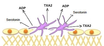 |
| 5 | As a result, platelets nearby are also activated, and the membrane glycoprotein receptor GPIIb/IIIa becomes active on their surface. | 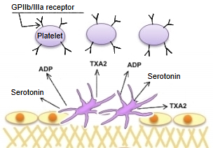 |
| 6 | vWF and fibrinogen act as bridges that join the platelets to one another. | 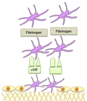 |
| 7 | The blood flow tends to pull these bonds apart (shear stress). | 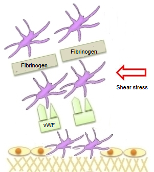 |
| 8 | Platelets activated by the shear stress release TXA2 and fibrinogen, which activate the receptors of other platelets and strengthen the bonds between them (platelet aggregation, also called secondary aggregation). | 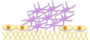 |
What a platelet contains
A platelet has no nucleus, but it does contain organelles such as granules and mitochondria, together with glycogen and systems of membranes and fibres.
(no nucleus)
- Granules : alpha granules, dense granules, glycogen granules
- Mitochondria : produce energy
- Open canalicular system : channels from the surface; the route by which granule contents are released
- Dense tubular system : stores calcium ions; makes thromboxane A2
- Cytoskeleton : microtubules and microfilaments
What the granules contain depends on the type of granule.
| Alpha granules | ||
| 1 | von Willebrand factor (vWF) | Binds to the collagen exposed beneath damaged endothelium. Platelets stick to it and release ADP and other substances, which draw in more platelets to form a platelet plug. |
| 2 | Platelet factor 4 (PF4) | A protein involved in blood clotting; by binding heparin (a polysaccharide) it blocks heparin's anticoagulant action. |
| 3 | Platelet-derived growth factor (PDGF) | Regulates the migration and multiplication of mesenchymal cells such as fibroblasts. |
| 4 | Thrombospondin (TSP) | A protein that binds heparin, fibrinogen, calcium ions and other substances and takes part in clotting. |
| 5 | Fibronectin | A glycoprotein that binds collagen, fibrin and other substances and takes part in clotting. |
| 6 | Fibrinogen | A plasma protein; it links platelets to one another and is converted to fibrin in clotting. |
| Dense granules | ||
| 1 | Serotonin | A monoamine neurotransmitter made from the essential amino acid tryptophan; it narrows injured vessels and activates platelets. |
| 2 | Adenosine diphosphate (ADP) | Activates and recruits other platelets. |
| 3 | Adenosine triphosphate (ATP) | A source of energy: energy is released when ATP is changed to ADP. |
| 4 | Calcium ions | Needed for platelet activation and for the clotting cascade. |
Reference websites
[Wikipedia]
· Platelet, Wikipedia.
· Megakaryocyte, Wikipedia.
Related terms
References and sources
- Further reading: Platelet, Wikipedia.
- Further reading: Megakaryocyte, Wikipedia.
- Federative International Programme for Anatomical Terminology (FIPAT). Terminologia Anatomica, 2nd ed. (TA2), 2019.