Splanchnic nerves (Latin: Nervi splanchnici)
Also called the visceral nerves
Overview
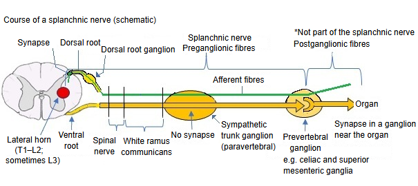
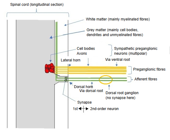
Structure at the origin
· The cell bodies (multipolar neurons) of the sympathetic efferent fibres that make up the splanchnic nerves lie in the lateral horn of the grey matter of the spinal cord (close to its border with the white matter), from T1 to L2 (sometimes L3).
· Their axons (efferent fibres) run in this order: ventral root ⇒ spinal nerve ⇒ white ramus communicans, and so reach the sympathetic trunk.
· The afferent fibres that travel with the efferent fibres pass through the dorsal root ganglion without synapsing and enter the dorsal root.
· They enter the spinal cord through the dorsal root and synapse in the dorsal horn with second-order neurons, whose fibres ascend in the white matter. (The afferent neuron, with its cell body in the dorsal root ganglion, is the first-order neuron.)
Types
By position, splanchnic nerves are grouped into thoracic, lumbar and pelvic splanchnic nerves.
splanchnic
nerves
- Thoracic splanchnic nerves : greater, lesser and least splanchnic nerves (sympathetic).
- Lumbar splanchnic nerves : lumbar splanchnic nerves (sympathetic).
- Pelvic splanchnic nerves : pelvic splanchnic nerves (parasympathetic).
| Name | Origin | Sympathetic trunk ganglion | In the ganglion | Fibres | Synapse | Main distribution | |
|---|---|---|---|---|---|---|---|
| 1 | Greater splanchnic nerve | T5–T9, lateral horn | Thoracic sympathetic trunk ganglia | Pass through | Sympathetic preganglionic fibres (mainly) + visceral afferent fibres | Celiac ganglion | Stomach, liver, spleen, pancreas, proximal small intestine |
| 2 | Lesser splanchnic nerve | T10–T11, lateral horn | Superior mesenteric ganglion | Small intestine, proximal colon | |||
| 3 | Least splanchnic nerve | T12, lateral horn | Renal ganglia and others | Kidney, suprarenal gland | |||
| 4 | Lumbar splanchnic nerves | L1–L2, lateral horn | Lumbar sympathetic trunk ganglia | Inferior mesenteric ganglion, pelvic plexuses | Distal colon, pelvic organs | ||
| 5 | Pelvic splanchnic nerves | S2–S4 | Not involved (parasympathetic fibres) | Parasympathetic preganglionic fibres (mainly) + visceral afferent fibres | Ganglia near the target organ or in its wall | Distal rectum, urinary bladder, genital organs, lower digestive tract | |
Sources differ on the segments of origin, for example T5–T9 or T5–T10 for the greater splanchnic nerve and T9–T10 or T10–T11 for the lesser splanchnic nerve.
Related terms
- Greater splanchnic nerve
- Lesser splanchnic nerve
- Least splanchnic nerve
- Lumbar splanchnic nerve
- Pelvic splanchnic nerves
- Sympathetic trunk
- Visceral afferent nerve
References and sources
- Further reading: Splanchnic nerves, Wikipedia.
- Federative International Programme for Anatomical Terminology (FIPAT). Terminologia Anatomica, 2nd ed. (TA2), 2019.