Gastrointestinal tract (Latin: tractus alimentarius)
Also called the alimentary canal, digestive tract
Overview
It is made up of the following parts.
intestinal
tract
- Oral cavity : the entrance of the tract; chews food, mixes it with saliva and prepares it for swallowing; digestion of starch begins.
- Pharynx : the food passage behind the oral cavity, also part of the respiratory system; it guides food into the esophagus.
- Esophagus : where the typical tube-shaped tract begins; a muscular organ that moves food on by peristalsis.
- Stomach : the widest part; stores and churns the contents (mechanical digestion) and digests them chemically with gastric juice.
- Small intestine : part of the intestine.
- Duodenum : neutralizes the contents with pancreatic juice and bile; full digestion begins.
- Jejunum : well-developed circular folds; the main part of the small intestine, the centre of nutrient absorption.
- Ileum : absorbs bile acids and vitamin B12; rich in lymphoid tissue.
- Large intestine : part of the intestine.
- Cecum : the contents pass into it from the ileocecal junction; important for immunity; the appendix hangs from it.
- Colon : absorbs water and electrolytes and forms the feces; three taeniae coli and the haustra.
- Rectum : stores the formed feces for a time.
- Anal canal : the last part of the tract; controls defecation with the internal (involuntary) and external (voluntary) anal sphincters.
The liver, gallbladder and pancreas are accessory digestive organs: they lie outside the tube and empty their secretions into the duodenum. The small and large intestines together form the intestine, and the pharynx is shared with the respiratory tract. Sources differ on the exact scope of the term: some texts start the tract at the mouth, others count only the parts from the stomach (or esophagus) to the anus.
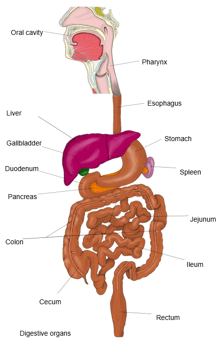
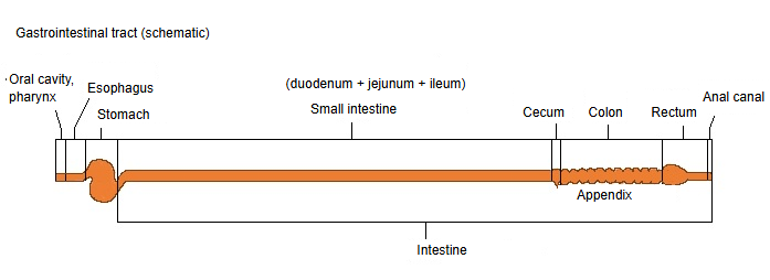
Comparison tables
Size
The table gives a simple overview of the length, inner diameter and wall thickness of each part (typical adult values; sources differ).
| 1 | 2 | 3 | 4 | 5 | 6 | 7 | 8 | 9 | 10 | 11 | |
| Small intestine | Large intestine | ||||||||||
| Oral cavity | Pharynx | Esophagus | Stomach | Duodenum | Jejunum | Ileum | Cecum | Colon | Rectum | Anal canal | |
| Length (cm) | a few cm | 12–14 | 25 | 25–30 | 25 | 250 | 350 | 6–8 | 150 | 12–15 | 3–4 |
| Inner diameter (cm) | variable | variable | 2–3 | 3–5 | 3–4 | 2.5–3 | 2–3 | 6–7 | 5–6 | 4–5 | 2–3 |
| Wall thickness (mm) | varies greatly | 2–4 | 2–3 | 5–7 | 2–3 | 2–3 | 1.5–2.5 | 2–3 | 2–3 | 3–4 | 2–3 |
Layers of the wall
The table gives a simple overview of how the layers of the wall differ between the parts.
| Part | Epithelium | Muscularis mucosae | Submucosa | Muscular layer | Outer layer | Typical features | |
|---|---|---|---|---|---|---|---|
| 1 | Oral cavity | Stratified squamous | Absent | Well developed | Mainly skeletal muscle | Adventitia | Unusual for the tract; close to the skin |
| 2 | Pharynx | Absent | Well developed | Skeletal muscle | Adventitia | Shared with the airway | |
| 3 | Esophagus | Distinct | Well developed | Upper: skeletal; middle: mixed; lower: smooth muscle | Adventitia (lowest part: serosa) | Resists friction | |
| 4 | Stomach | Simple columnar | Well developed | Three layers (with an oblique layer) | Serosa | Gastric glands, gastric folds | |
| 5 | Duodenum | Duodenal glands | Two layers (inner circular + outer longitudinal) | Serosa / adventitia | Neutralizes gastric acid | ||
| 6 | Jejunum | Moderate | Serosa | Circular folds most developed | |||
| 7 | Ileum | Peyer's patches | Serosa | Immune function | |||
| 8 | Cecum | Well developed | Inner circular + outer longitudinal (= taeniae coli) | Serosa | Much lymphoid tissue | ||
| 9 | Colon | Well developed | Serosa / adventitia | Haustra | |||
| 10 | Rectum | Well developed | Two layers (taeniae end) | Mainly adventitia | Ampulla | ||
| 11 | Anal canal | Upper: simple columnar; lower: stratified squamous | Unclear | Well developed | Internal anal sphincter (sphincter) | Adventitia | Change of epithelium (pectinate line) |
From the esophagus downwards the wall has the same basic layers: mucosa, submucosa, muscular layer and an outer serosa or adventitia. The muscle is skeletal in the mouth, pharynx and upper esophagus and smooth further down. The lining changes from stratified squamous epithelium (mouth to esophagus) to simple columnar epithelium (stomach to rectum) and back to stratified squamous in the lower anal canal.
Functions
The table gives a simple overview of the work of each part.
| Part | Main functions | Key points | |
|---|---|---|---|
| 1 | Oral cavity | Intake, chewing, first digestion | Chewing, secretion of saliva, start of carbohydrate digestion by amylase |
| 2 | Pharynx | Swallowing, passage | Sends the bolus into the esophagus (almost no digestion) |
| 3 | Esophagus | Transport | Moves the bolus on by peristalsis; prevents reflux |
| 4 | Stomach | Storage, churning, chemical digestion | Gastric acid and pepsin; digestion of protein; killing microbes |
| 5 | Duodenum | Neutralization, regulation of digestion | Neutralizes gastric acid; mixes in bile and pancreatic juice; the hub of digestion |
| 6 | Jejunum | Main absorption | Absorbs most of the sugars, amino acids and fats |
| 7 | Ileum | Selective absorption, immunity | Absorbs bile acids and vitamin B12; immune surveillance |
| 8 | Cecum | Fermentation, immunity | Activity of the gut bacteria; rich in lymphoid tissue |
| 9 | Colon | Recovering water, forming feces | Absorbs water and electrolytes; concentrates the feces |
| 10 | Rectum | Storage, defecation reflex | Stores feces; starts the defecation reflex |
| 11 | Anal canal | Control of defecation | Controls defecation with the internal (involuntary) and external (voluntary) anal sphincters |
Related terms
References and sources
- Further reading: Gastrointestinal tract, Wikipedia.
- Further reading: Gastrointestinal wall, Wikipedia.
- Federative International Programme for Anatomical Terminology (FIPAT). Terminologia Anatomica, 2nd ed. (TA2), 2019.