Mesangial cell (Latin: mesangiocytus)
Overview
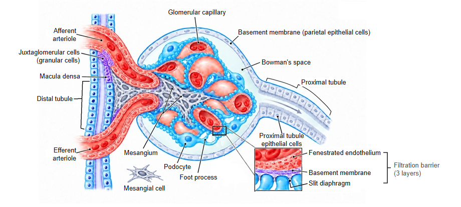
· A mesangial cell is a cell of the mesangium, the tissue that fills the spaces between the glomerular capillaries inside the glomerulus of the renal corpuscle and supports them. The spaces between the cells are filled by the mesangial matrix, which the cells produce themselves. The renal corpuscle has three components: the glomerulus, its supporting mesangium, and the two-layered epithelial glomerular capsule (Bowman's capsule) that encloses them.
· There are two groups: intraglomerular mesangial cells, between the capillary loops inside the glomerulus, and extraglomerular mesangial cells, which lie between the afferent arteriole and the efferent arteriole at the vascular pole. "Mesangial cell" on its own usually means the intraglomerular type. The two groups are continuous at the vascular pole.
· The cells are irregular in shape, and their processes contain actin, myosin and related proteins, so they can contract. They support the capillaries from the inside and are thought to help regulate capillary blood flow and the surface area available for filtration.
· They are separated from the capillary lumen only by the fenestrated endothelial cells, so they are exposed to large molecules from the blood. Mesangial cells take up material that collects in the glomerular basement membrane and the mesangium and clear it away, keeping the filter that forms primary urine clean.
· Mesangial proliferative glomerulonephritis, which includes IgA nephropathy, is a form of nephritis in which the mesangial cells or the mesangial matrix increase.
Reference websites
[Wikipedia]
· Mesangial cell, Wikipedia.
· Mesangium, Wikipedia.
[Websites with illustrations]
· Renal corpuscle (Digital Histology)
· Renal corpuscle (histology) (SIU School of Medicine)
· Glomeruli: histology (kidneypathology.com)
· Renal corpuscle (diagram) (Wikimedia Commons)

Related terms
- Mesangium
- Mesangial matrix
- Extraglomerular mesangial cell
- Glomerular capillary
- Glomerular basement membrane
- Juxtaglomerular apparatus
- Endothelial cell
References and sources
- Further reading: Mesangial cell, Wikipedia.
- Further reading: Mesangium, Wikipedia.
- Federative International Programme for Anatomical Terminology (FIPAT). Terminologia Anatomica, 2nd ed. (TA2), 2019.