Vascular pole (Latin: polus vascularis)
Overview
· The vascular pole lies exactly opposite the urinary pole, where the renal tubule begins.
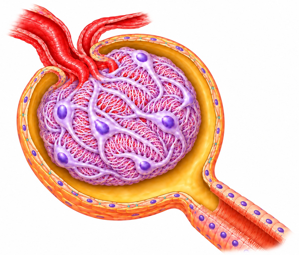
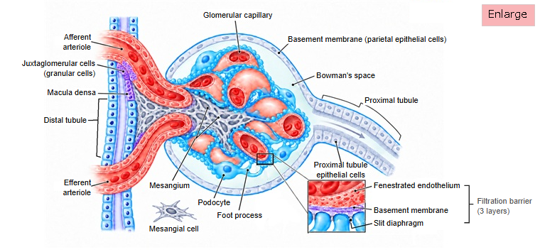
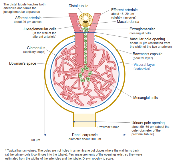
· At the vascular pole the wall of Bowman's capsule folds back: the outer wall (parietal layer) turns here into the inner wall (visceral layer) that covers the capillaries of the glomerulus.
· The afferent arteriole, a branch of an interlobular artery, enters the renal corpuscle at the vascular pole and divides into the glomerular capillaries. These capillaries join again to form a single efferent arteriole, which leaves through the same pole.
· Near the vascular pole, the renin-producing juxtaglomerular cells lie in the wall of the afferent arteriole, and the macula densa in the wall of the distal tubule of the same nephron touches them. These are parts of the juxtaglomerular apparatus, which regulates the work of the glomerulus.
· In histological sections of a renal corpuscle, the side where arterioles enter and leave and a macula densa is seen is the vascular pole; the side where the proximal tubule leaves is the urinary pole. These features tell the two poles apart.
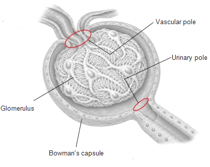
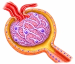 Renal corpuscle (section)
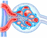 Renal corpuscle (section)
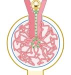 Renal corpuscle, diagram (section)
Reference websites
[Wikipedia]
· Renal corpuscle, Wikipedia.
· Juxtaglomerular apparatus, Wikipedia.
[Websites with illustrations]
· Renal corpuscle, vascular pole (micrograph) (SIU School of Medicine histology)
· Renal corpuscle (micrograph) (SIU School of Medicine histology)
· Renal corpuscle (histology image) (Tufts University histology images)
· Tubules and collecting system (Digital Histology)
Related terms
- Distal tubule
- Glomerular capillary
- Glomerular basement membrane
- Podocyte
- Visceral layer of glomerular capsule
- Parietal layer of glomerular capsule
- Capsular space
- Urinary pole
- Macula densa
- Juxtaglomerular apparatus
- Juxtaglomerular cells
- Mesangium
- Interlobular arteries of kidney (cortical radiate arteries)
- Kidney
References and sources
- Further reading: Renal corpuscle, Wikipedia.
- Further reading: Juxtaglomerular apparatus, Wikipedia.
- Federative International Programme for Anatomical Terminology (FIPAT). Terminologia Anatomica, 2nd ed. (TA2), 2019.