Mesangium (Latin: mesangium)
Overview
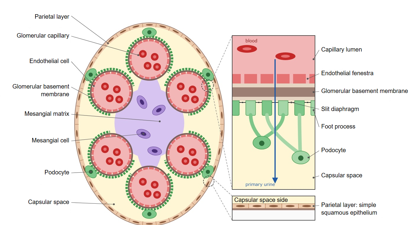
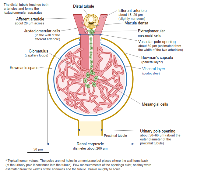
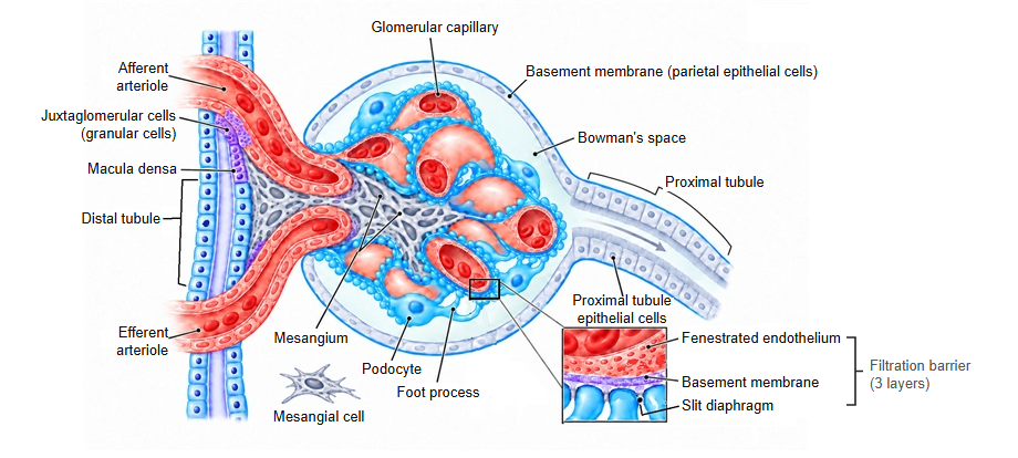
· There are two groups of mesangial cells. Intraglomerular mesangial cells lie between the capillaries of the glomerulus. Extraglomerular mesangial cells (lacis or Goormaghtigh cells) lie between the afferent arteriole and the efferent arteriole at the vascular pole; they are part of the juxtaglomerular apparatus.
· Mesangial cells support the glomerular capillaries, produce the mesangial matrix, and take up foreign material and protein that collect in the glomerular basement membrane and the mesangium, clearing them away and keeping the filter clear.
· The cells can contract, and they are thought to affect blood flow through the capillaries and the surface area available for filtration.
· Mesangial proliferative glomerulonephritis is a disease in which the mesangial cells or the mesangial matrix increase.
The chart below shows the parts of the mesangium in a simple form.
- Mesangial cells
- Intraglomerular mesangial cells : support the capillaries, make the matrix and take up foreign material
- Extraglomerular mesangial cells : at the vascular pole; part of the juxtaglomerular apparatus (Goormaghtigh cells)
- Mesangial matrix : the matrix that fills the spaces between the mesangial cells; for example type IV collagen, type V collagen, laminin, fibronectin and proteoglycans
Reference websites
[Wikipedia]
· Mesangium, Wikipedia.
· Mesangial cell, Wikipedia.
[Websites with illustrations]
· Renal corpuscle (Digital Histology)
· Renal corpuscle (histology) (SIU School of Medicine)
· Glomeruli: histology (kidneypathology.com)
· Renal corpuscle (diagram) (Wikimedia Commons)

Related terms
- Visceral layer of glomerular capsule
- Parietal layer of glomerular capsule
- Capsular space
- Urinary pole
- Glomerular capillary
- Glomerular basement membrane
- Juxtaglomerular apparatus
- Juxtaglomerular cells
- Vascular pole
- Podocyte
- Kidney
References and sources
- Further reading: Mesangium, Wikipedia.
- Further reading: Mesangial cell, Wikipedia.
- Further reading: Glomerulus (kidney), Wikipedia.
- Federative International Programme for Anatomical Terminology (FIPAT). Terminologia Anatomica, 2nd ed. (TA2), 2019.