Latin: musculus popliteus
Overview
- One of the flexor group of the leg (among the “others” besides the triceps surae); originally a deep flexor of the leg.
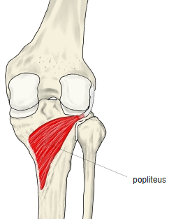
- A flat, triangular muscle, thicker in its middle, forming the floor of the popliteal fossa (the hollow at the back of the knee).
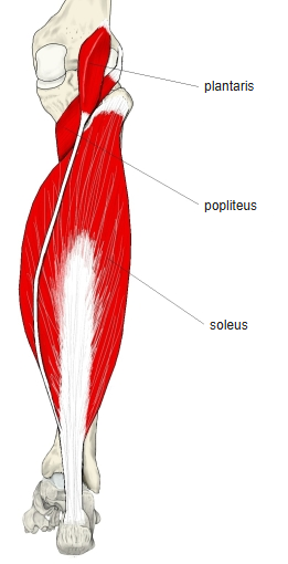
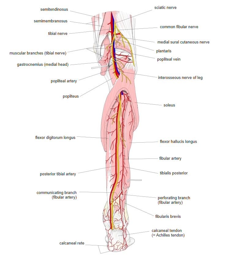
- Covered by the heads of the gastrocnemius and by the popliteal vessels and nerves.
- The only muscle of the back of the leg that acts on the knee but not on the ankle.
- In non-mammals it connects the tibia and fibula; in mammals it developed in close relation to the knee joint.
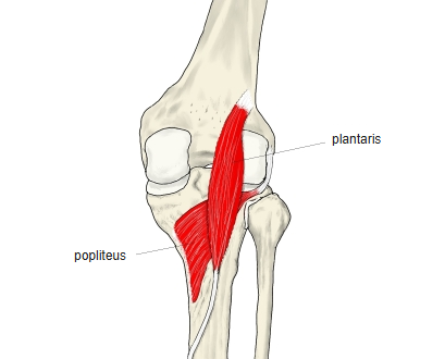
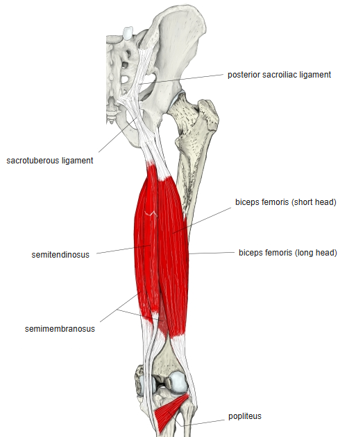
- Continuous in places with the tibialis posterior, plantaris, semimembranosus, biceps femoris (short head) and soleus.
Position in the classification
Lower Limb › Leg › Flexor group › Others
Illustrations
Origin / insertion
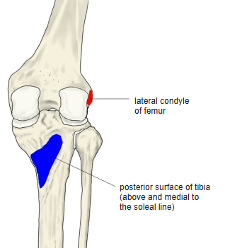
- Origin: outer surface of the lateral condyle of the femur (below the lateral collateral ligament), by a rounded tendon; also the lateral collateral ligament and the capsule of the knee joint
- The origin lies inside the joint capsule; the tendon runs between the lateral meniscus and the lateral collateral ligament, and the arcuate popliteal ligament passes over it.
- Part of the muscle attaches to the lateral meniscus.
- The fibres run downward and medially.
- Insertion: posterior surface of the tibia, above the soleal line
Action
- Flexes the knee and rotates the leg (tibia) medially
- Foot on the ground: rotates the femur laterally on the tibia and so “unlocks” the extended knee at the start of flexion
- Pulls the lateral meniscus backward early in flexion, so it is not crushed between femur and tibia
- Used in walking, sitting down and standing up
Innervation
- Tibial nerve (L4–S1; English sources: L5–S1)
Blood supply
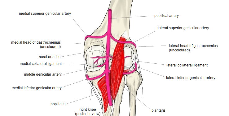
- Medial inferior genicular artery
- Other branches of the popliteal artery
Anatomical variations
- An extra head may arise from the sesamoid bone in the lateral head of the gastrocnemius
- Rarely a small extra muscle, the popliteus minor, runs from the femur (medial to the plantaris) to the posterior ligament of the knee
- Peroneotibialis (about 14%): from the head of the fibula to the tibia, beneath the popliteus
- A small sesamoid bone, the cyamella, is rarely found in its tendon
Related terms
- Popliteal fossa
- Knee joint
- Lateral condyle of femur
- Lateral collateral ligament
- Lateral meniscus
- Arcuate popliteal ligament
- Soleal line
- Plantaris
- Semimembranosus
- Tibial nerve
- Popliteal artery
- Medial inferior genicular artery
References and sources
- Further reading: Popliteus muscle, Wikipedia.