Latin: concha nasalis inferior
Overview
- A paired, thin, curled bone attached to the lateral wall of the nasal cavity. Unlike the superior and middle conchae, which belong to the ethmoid, it is an independent bone.
- It is the largest of the three conchae and separates the middle nasal meatus above from the inferior nasal meatus below; the nasolacrimal canal opens into the inferior meatus beneath it.
- It develops from the lower part of the cartilaginous nasal capsule and begins to ossify around the fifth to sixth fetal month.
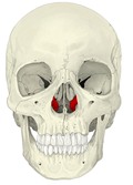
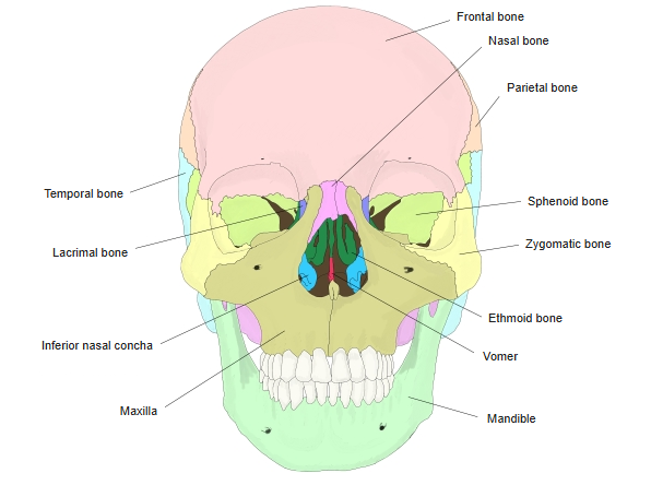
Skull (anterior) 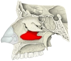
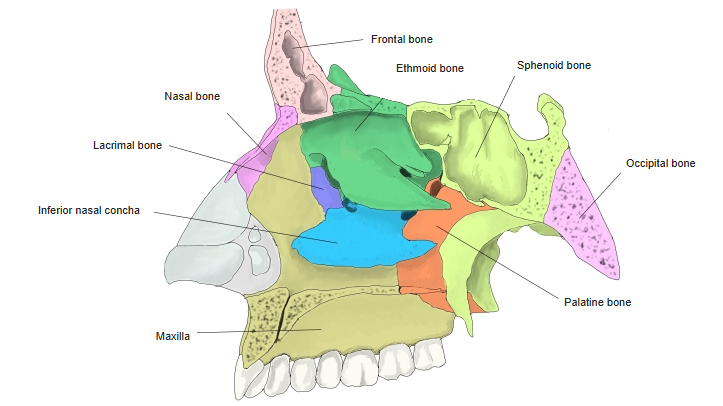
Lateral wall of the nasal cavity 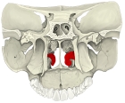
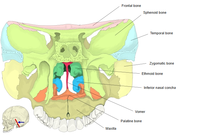
Coronal section (from behind) 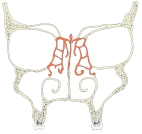
Coronal section of the nasal cavity
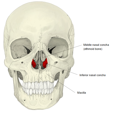
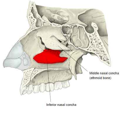
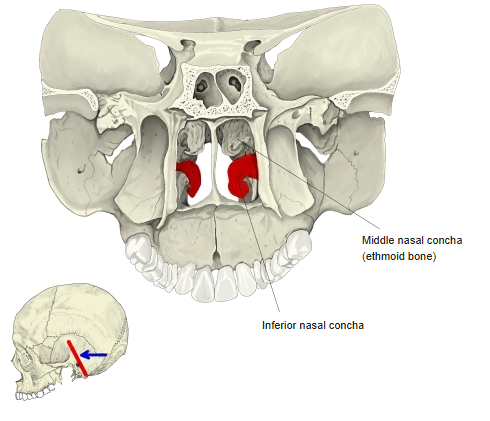
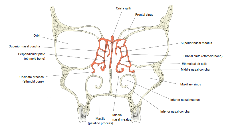
- Bones of the skull
- Neurocranium (cranial bones)
- Frontal bone
- Parietal bone (2)
- Occipital bone
- Temporal bone (2)
- Sphenoid bone
- Ethmoid bone
- Viscerocranium (facial bones)
- Nasal bone (2)
- Vomer
- Lacrimal bone (2)
- Inferior nasal concha (2)
- Maxilla (2)
- Zygomatic bone (2)
- Palatine bone (2)
- Mandible
- Hyoid bone
- Neurocranium (cranial bones)
Illustrations
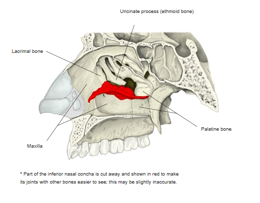
Articulations
| # | Bone | Joint |
|---|---|---|
| 1 | Maxilla | Conchal crest; maxillary process at the maxillary hiatus |
| 2 | Lacrimal bone | Lacrimal process |
| 3 | Ethmoid bone | Ethmoidal process – uncinate process |
| 4 | Palatine bone | Conchal crest of the perpendicular plate |
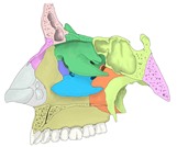
Lateral wall of the nasal cavity (coloured) 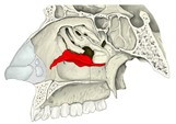
Neighbouring bones 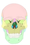
Skull (anterior, coloured) 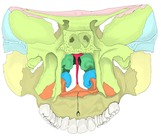
Coronal section (coloured)
Muscle attachments
- Origin of: none
- Insertion of: none
No muscles attach to the inferior nasal concha.
Borders and processes
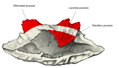
- Superior border – thin and irregular; it is attached to the lateral wall of the nasal cavity and carries three processes:
- Lacrimal process – projects upwards in front and meets the lacrimal bone, completing the nasolacrimal canal.
- Ethmoidal process – projects upwards behind and meets the uncinate process of the ethmoid.
- Maxillary process – curves downwards and laterally, partly closing the maxillary hiatus.
- Inferior border – free, thick and spongy, curling slightly inwards.
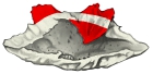
Processes (medial view) 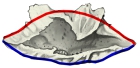
Borders
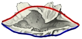
Surfaces
- Medial surface – convex and rough, facing the nasal septum.
- Lateral surface – concave, facing the inferior nasal meatus.
References and sources
- Further reading: Inferior nasal concha, Wikipedia.