Nasal cavity (Latin: cavitas nasi)
- Overview
- Bones and cartilages
- Nasal mucosa
- Openings
- Nerves and vessels
- Terms related to the nasal cavity
- Reference websites
- (Wikipedia, websites with illustrations)
- Where it is found
- Related terms
Overview
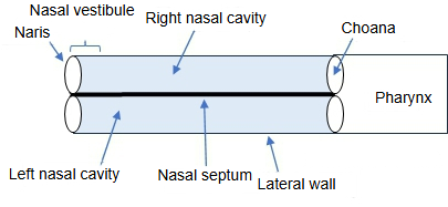
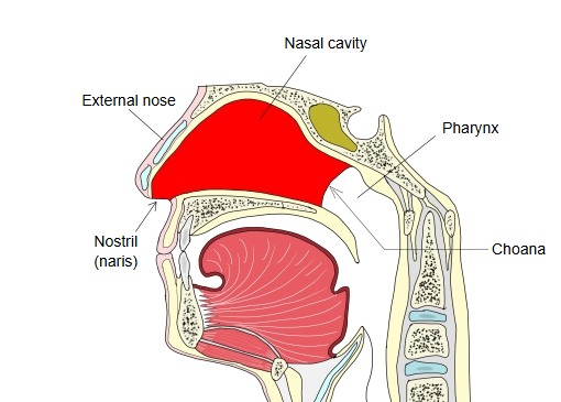
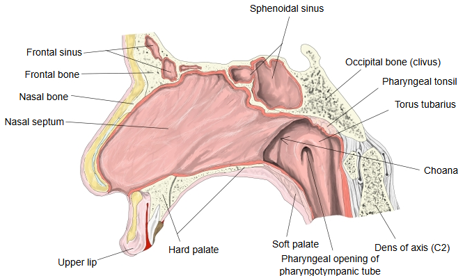
The nasal cavity can be divided into the nasal vestibule, the narrow front part just inside the nostril (lined with skin and hairs), and the nasal cavity proper behind it.
The roof is narrow and formed mainly by the cribriform plate of the ethmoid bone; the floor is the hard palate. Behind, the cavity opens through the choanae into the nasal part of the pharynx (nasopharynx).
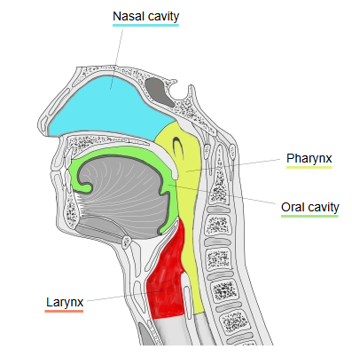
- Respiratory tract
- Nasal cavity : from the nostrils to the choanae; divided into right and left halves by the nasal septum and lined with mucosa.
- Pharynx : made of three parts (nasal, oral and laryngeal) and lined with mucosa; also part of the digestive system.
- Larynx : a funnel-shaped tube at the level of the 4th-7th cervical vertebrae; its framework is made of cartilage.
- Trachea : a tube about 10 cm long and 20-25 mm across; divides at the level of the 4th-5th thoracic vertebrae.
- Bronchi : on both sides they branch from the trachea, reach the hilum of the lung and divide into many bronchial branches.
- Lungs : right lung (upper, middle and lower lobes), left lung (upper and lower lobes); gas exchange (external respiration) takes place in the alveoli.
The respiratory system: the respiratory tract (the air passage) and the lungs
Nasal cavity to bronchi = the respiratory tract (the passage for air).
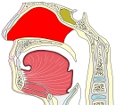 Nasal cavity
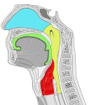 Nasal cavity to larynx
Bones and cartilages
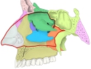
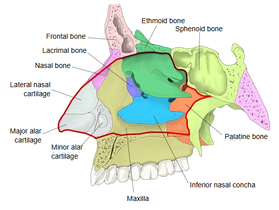
The bones and cartilages that form the nasal cavity are listed below.
| Bones | Nasal bone, palatine bone, ethmoid bone, maxilla, inferior nasal concha, frontal bone, lacrimal bone, sphenoid bone, vomer |
| Cartilages | Lateral nasal cartilage, major alar cartilage, minor alar cartilages, septal cartilage (in the front of the nose) |
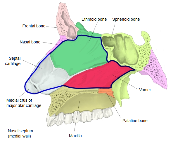
The nasal septum is formed by the septal cartilage in front, the perpendicular plate of the ethmoid above and behind, and the vomer below and behind.
 Lateral wall: bones and cartilages
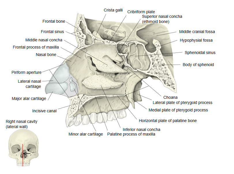
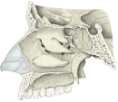 Lateral wall (bone)
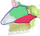 Medial wall: nasal septum
Nasal mucosa
[Mucosa]
The inside of the nasal cavity is covered by a fairly thick mucous membrane (the nasal mucosa), so the open space is much narrower than the bony cavity. The mucosa has two parts, the respiratory region and the olfactory region. See Nasal mucosa for details.
- Respiratory region : most of the mucosa of the nasal cavity; rich in blood vessels and pale red. It warms and moistens the air (part of the respiratory system).
- Olfactory region : a small yellowish area in the upper part of the cavity (the roof and the upper septum) containing the olfactory cells (the organ of smell).
[Lateral wall]
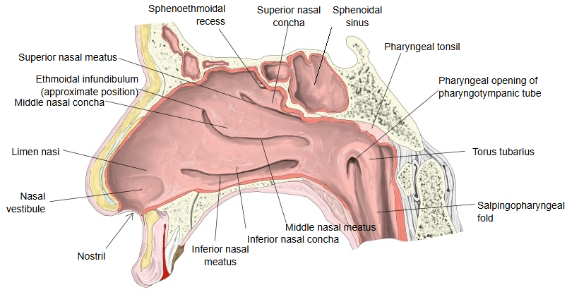
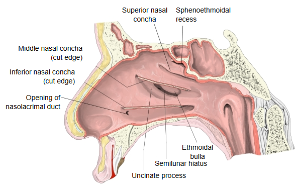
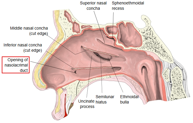
Unlike the smooth medial wall (the septum), the lateral wall has a complex shape. The ethmoidal bulla, uncinate process, semilunar hiatus and ethmoidal infundibulum lie in the middle meatus, hidden under the middle concha.
· Nasal conchae: three shelves running from front to back; the lower edge of each curls downwards and forms an air passage (a nasal meatus) under it.
conchae
- Superior nasal concha : above the superior nasal meatusWhen the back part of the superior concha is divided into an upper and a lower part, the upper one is called the supreme nasal concha.
- Middle nasal concha : above the middle nasal meatus
- Inferior nasal concha : above the inferior nasal meatus
· Sphenoethmoidal recess: a narrow space above and behind the superior nasal concha and in front of the body of the sphenoid, between the superior concha and the nasal septum.
· Ethmoidal bulla: a rounded bulge on the lateral wall of the middle meatus, under the middle nasal concha, formed by ethmoidal air cells of the ethmoidal labyrinth; it is regarded as a rudimentary concha.
· Uncinate process: a projecting plate in front of the semilunar hiatus. The hiatus lies between the uncinate process and the ethmoidal bulla.
· Semilunar hiatus: the curved slit between the ethmoidal bulla and the uncinate process.
· Ethmoidal infundibulum: at its front upper end the semilunar hiatus narrows and continues into this tube-like channel in the front part of the ethmoidal labyrinth; the frontal sinus usually drains through it.
[Others]
· Common nasal meatus: the inner edges of the conchae do not reach the nasal septum, so on each side of the septum the three meatuses are joined by a common narrow vertical space. Behind, it continues to the choana.
 Mucosa of the lateral wall (1)
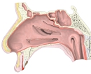 Mucosa of the lateral wall (2)
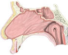 Mucosa of the nasal septum
Openings
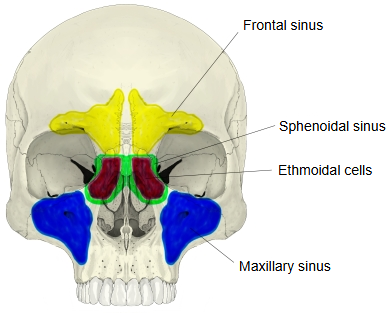
- Maxillary sinus : opens mainly through the semilunar hiatus into the middle nasal meatus; a small accessory opening is often present behind the semilunar hiatus.
- Frontal sinus : opens at the front upper end of the ethmoidal infundibulum into the middle nasal meatus.
- Ethmoidal cells : the anterior and middle cells open into the upper part of the middle nasal meatus (the middle cells on or above the ethmoidal bulla); the posterior cells open into the superior nasal meatus.
- Sphenoidal sinus : opens into the sphenoethmoidal recess.
· The nasolacrimal duct opens into the front of the inferior nasal meatus, about 30-35 mm behind the nostril.
 Right lateral wall (conchae removed)
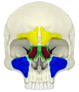 Paranasal sinuses
Nerves and vessels
[Nerves]
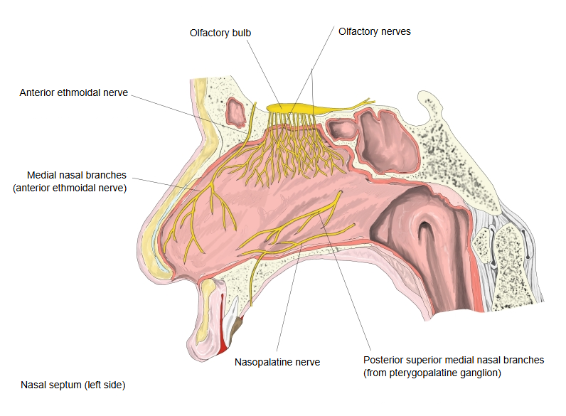
| Nasal mucosa: respiratory region | · Anterior ethmoidal nerve: a branch of the nasociliary nerve, one of the branches of the ophthalmic nerve (V1); supplies the upper front part. · Nasal branches from the pterygopalatine ganglion, reached through the pterygopalatine nerves of the maxillary nerve (the 2nd division of the trigeminal nerve, V2), including the nasopalatine nerve; they supply most of the rest. |
| Nasal mucosa: olfactory region | · Olfactory nerves (cranial nerve I): carry smell from the olfactory region through the cribriform plate to the olfactory bulb. |
 Nerves of the nasal septum
[Vessels]
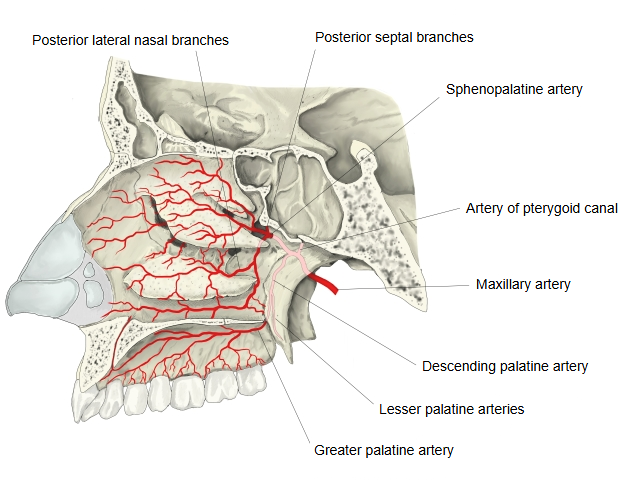
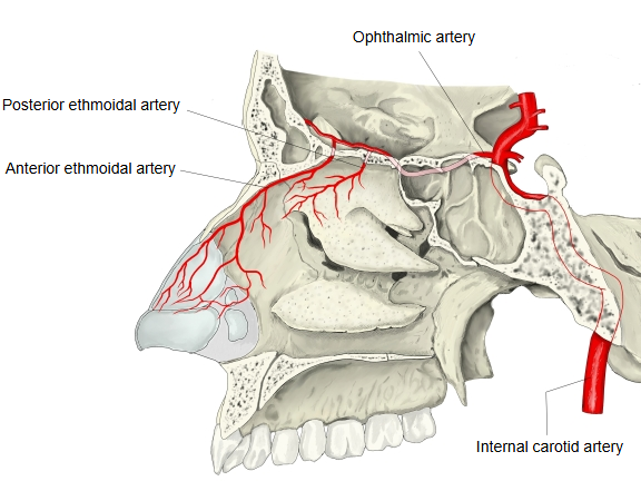
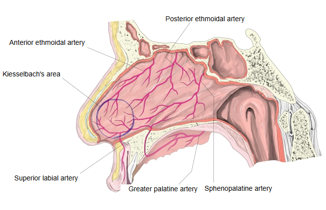
| Arteries | 1. Sphenopalatine artery (from the maxillary artery); main branches: the posterior lateral nasal branches and the posterior septal nasal branches. 2. Angular artery (from the facial artery). 3. Anterior ethmoidal artery (from the ophthalmic artery, a branch of the internal carotid artery). 4. Posterior ethmoidal artery (also from the ophthalmic artery). The greater palatine artery and further branches of the facial artery (the superior labial artery and the lateral nasal branches) also take part. |
| Veins | Run with the arteries and connect with the veins of the face, the orbit and the mouth. |
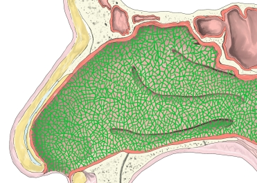
| Lymph vessels | Form a network in the lamina propria of the mucosa and drain mainly to the lymph nodes along the 2nd cervical vertebra (axis) and the greater horn of the hyoid bone: the deep cervical, retropharyngeal and submandibular lymph nodes. Some lymph also goes to the nodes near the parotid gland. |
These arteries meet on the lower front part of the nasal septum in Kiesselbach's area, the usual site of nosebleeds.
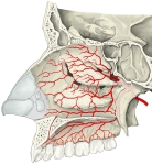 Arteries of the lateral wall (1)
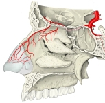 Arteries of the lateral wall (2)
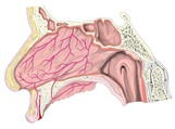 Left nasal cavity: arteries of the nasal septum
 Lymph vessels
Terms related to the nasal cavity
- Cavernous plexus of conchae
- Choana (= internal naris)
- Common nasal meatus
- Ethmoidal bulla
- Ethmoidal infundibulum
- Inferior nasal concha
- Inferior nasal meatus
- Kiesselbach's area
- Middle nasal concha
- Middle nasal meatus
- Naris (nostril)
- Nasal glands
- Nasal mucosa (olfactory region, respiratory region)
- Nasal septum
- Nasal vestibule
- Nasolacrimal duct
- Paranasal sinuses
- Semilunar hiatus
- Sphenoethmoidal recess
- Superior nasal concha
- Superior nasal meatus
- Uncinate process
- Vibrissae (nasal hairs)
Reference websites
[Wikipedia]
· Nasal cavity, Wikipedia.
· Paranasal sinuses, Wikipedia.
· Kiesselbach's plexus, Wikipedia.
[Websites with illustrations]
· The nasal cavity (TeachMeAnatomy)
· Nasal cavity: anatomy, structure, parts, blood supply (Kenhub)
· Lateral wall of the nasal cavity (Gray's Anatomy, fig. 855) (Wikimedia Commons)

Where it is found
Related terms
- Nasal septum
- Superior nasal concha
- Middle nasal concha
- Inferior nasal meatus
- Middle nasal meatus
- Piriform aperture
- Maxillary sinus
References and sources
- Further reading: Nasal cavity, Wikipedia.
- Further reading: Paranasal sinuses, Wikipedia.
- Further reading: Kiesselbach's plexus, Wikipedia.
- Federative International Programme for Anatomical Terminology (FIPAT). Terminologia Anatomica, 2nd ed. (TA2), 2019.