Latin: os lacrimale
Overview
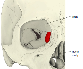
- A small, thin paired bone at the front of the medial wall of the orbit; it is the smallest and most fragile bone of the face.
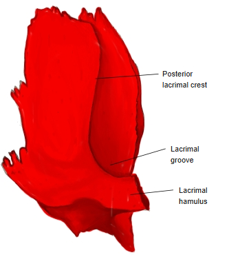
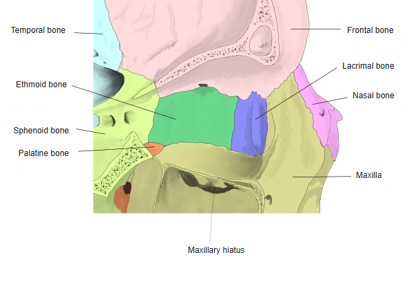
- Its lateral (orbital) surface forms part of the orbit and is divided by the vertical posterior lacrimal crest into a front part, the lacrimal groove (which, with the maxilla, forms the fossa for the lacrimal sac), and a back part. The crest ends below in the hook-like lacrimal hamulus.
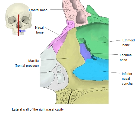
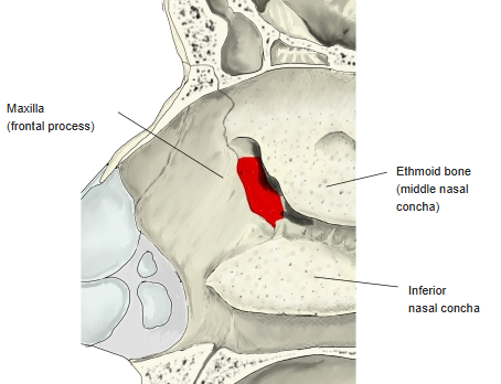
- Its medial (nasal) surface forms part of the lateral wall of the nasal cavity.
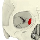
Right orbit 
Right lacrimal bone (lateral surface)
- Bones of the skull
- Neurocranium (cranial bones)
- Frontal bone
- Parietal bone (2)
- Occipital bone
- Temporal bone (2)
- Sphenoid bone
- Ethmoid bone
- Viscerocranium (facial bones)
- Nasal bone (2)
- Vomer
- Lacrimal bone (2)
- Inferior nasal concha (2)
- Maxilla (2)
- Zygomatic bone (2)
- Palatine bone (2)
- Mandible
- Hyoid bone
- Neurocranium (cranial bones)
Illustrations
Articulations
| # | Bone | Joint |
|---|---|---|
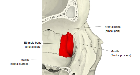
| 1 | Frontal bone | Orbital part |
| 2 | Ethmoid bone | Orbital plate |
| 3 | Maxilla | Frontal process and orbital surface |
| 4 | Inferior nasal concha | Lacrimal process |
Medial wall of the right orbit 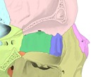
Medial wall of the orbit (coloured) 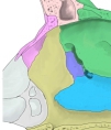
Lateral wall of the right nasal cavity 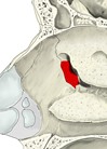
Lateral wall of the nasal cavity
Muscle attachments
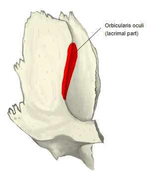
- Origin of: Orbicularis oculi (lacrimal part)
- Insertion of: none
The lacrimal part of orbicularis oculi arises from the posterior lacrimal crest.
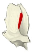
Muscle attachment
Borders
- Anterior border – joins the frontal process of the maxilla.
- Posterior border – joins the orbital plate of the ethmoid.
- Superior border – joins the orbital part of the frontal bone.
- Inferior border – joins the orbital surface of the maxilla; in front, the descending process meets the inferior nasal concha.
Development and variations
- It develops in membrane on the outer surface of the cartilaginous nasal capsule and begins to ossify around the third fetal month.
- It is occasionally absent; the lacrimal hamulus may be very small, and the bone sometimes has a hole.
References and sources
- Further reading: Lacrimal bone, Wikipedia.