Latin: os hyoideum
Overview
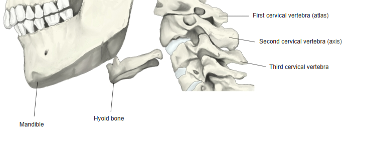
- A small U-shaped bone in the front of the neck, between the mandible and the larynx, at about the level of the third cervical vertebra.
- It does not articulate with any other bone; it is held in place by muscles and ligaments – the suprahyoid muscles above and the infrahyoid muscles below – and supports the root of the tongue.
- It consists of a body in front, a pair of greater horns projecting backwards and a pair of small lesser horns projecting upwards where the body meets the greater horns.
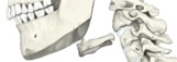
Position of the hyoid bone
- Bones of the skull
- Neurocranium (cranial bones)
- Frontal bone
- Parietal bone (2)
- Occipital bone
- Temporal bone (2)
- Sphenoid bone
- Ethmoid bone
- Viscerocranium (facial bones)
- Nasal bone (2)
- Vomer
- Lacrimal bone (2)
- Inferior nasal concha (2)
- Maxilla (2)
- Zygomatic bone (2)
- Palatine bone (2)
- Mandible
- Hyoid bone
- Neurocranium (cranial bones)
Illustrations
Ligaments
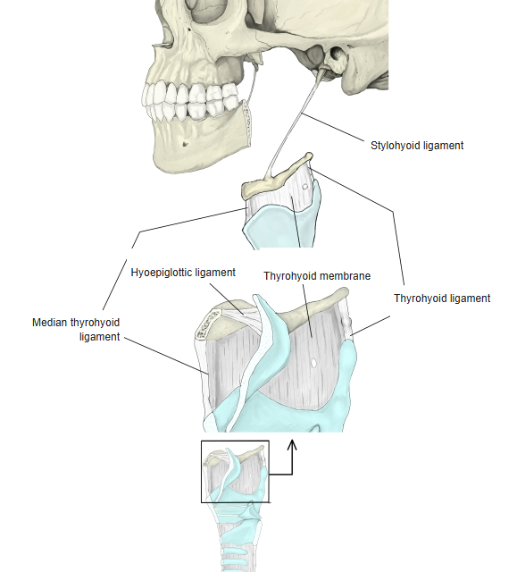
The following ligaments attach to the hyoid bone. The list is intended to be comprehensive but may omit minor ligaments.
| # | Ligament | Connects |
|---|---|---|
| 1 | Stylohyoid ligament | Styloid process of the temporal bone – lesser horn |
| 2 | Thyrohyoid membrane | Upper border of the thyroid cartilage – hyoid bone |
| 3 | Median thyrohyoid ligament | Thickened middle part of the thyrohyoid membrane |
| 4 | Lateral thyrohyoid ligament | Thickened back edge of the thyrohyoid membrane (superior horn of thyroid cartilage – tip of greater horn) |
| 5 | Hyoepiglottic ligament | Epiglottis – back of the body of the hyoid |
Ligaments of the hyoid bone
Muscle attachments
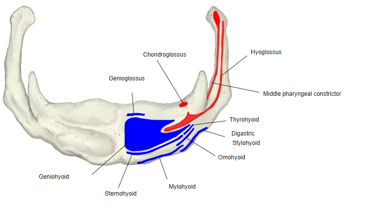
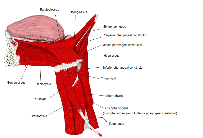
- Origin of: Hyoglossus, Chondroglossus, Middle pharyngeal constrictor, Genioglossus (lowest fibres)
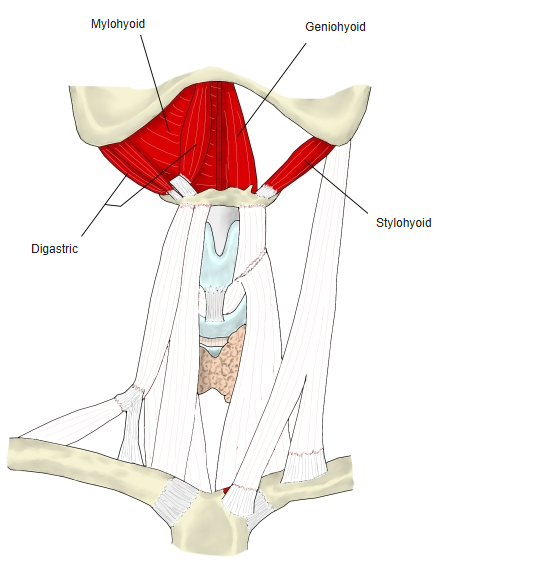
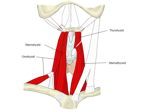
- Insertion of: Mylohyoid, Geniohyoid, Stylohyoid, Digastric (via a fibrous loop on the intermediate tendon), Sternohyoid, Omohyoid, Thyrohyoid
In the attachment figure, origins are shown in red and insertions in blue. The suprahyoid muscles raise the hyoid; the infrahyoid muscles lower it.
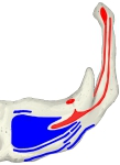
Muscle attachments 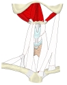
Suprahyoid muscles 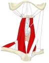
Infrahyoid muscles 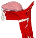
Muscles around the hyoid (lateral)
Parts
- Body – the quadrilateral front part; its anterior surface is convex and gives attachment to many muscles.
- Greater horns – project backwards from the sides of the body; their tips end in small tubercles.
- Lesser horns – small conical projections at the junction of the body and greater horns; the stylohyoid ligament attaches to their tips.
References and sources
- Further reading: Hyoid bone, Wikipedia.