Latin: axis · second cervical vertebra (C2)
Overview
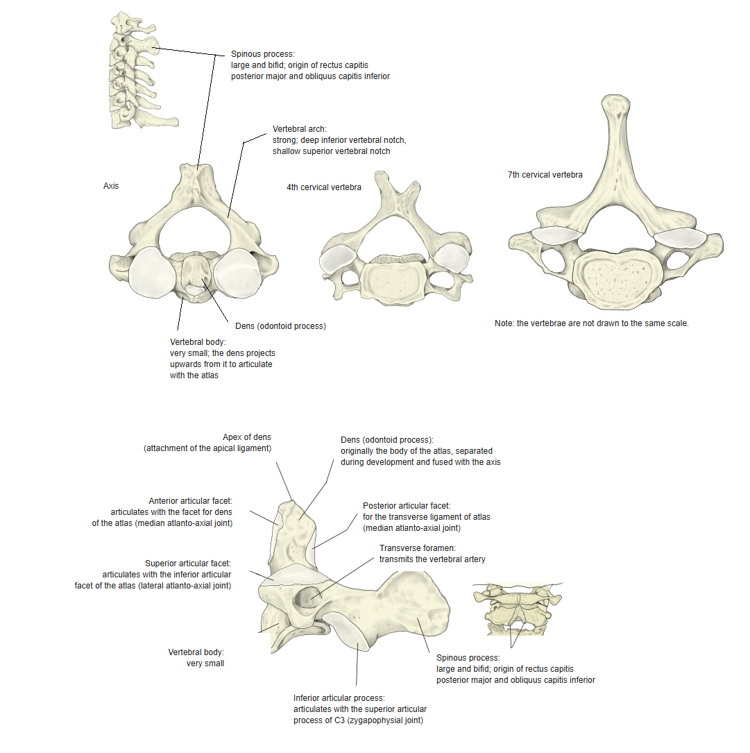
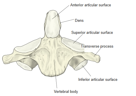
- The second cervical vertebra (C2). Its most characteristic feature is the dens (odontoid process), a tooth-like peg that projects upwards from its small body.
- The dens forms the pivot around which the atlas – and with it the head – rotates at the median atlanto-axial joint.
- Developmentally the dens is the body of the atlas, which separates from the atlas and fuses with the body of the axis.
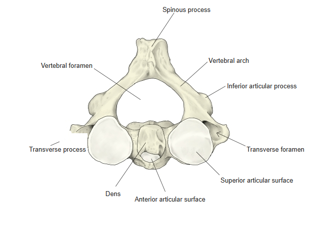
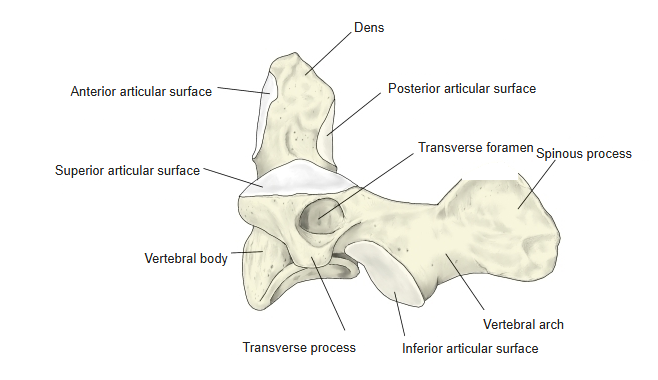
- The body of the axis is small compared with those of the lower cervical vertebrae; the pedicles are broad and strong, especially in front beneath the superior articular facets, and the laminae are thick. The spinous process is large and bifid, and is the first spinous process that can be felt below the occipital bone.
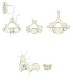
Axis (posterosuperior view) 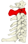
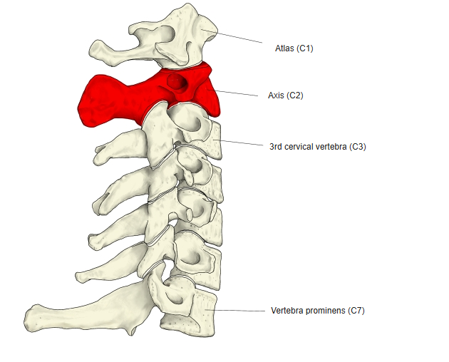
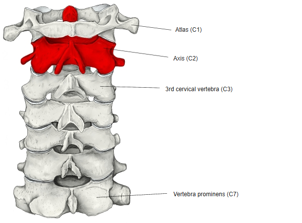
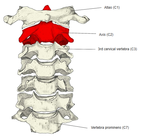
Cervical vertebrae (left lateral), axis highlighted
- Bones of the trunk
- Vertebral column
- Cervical vertebrae (7)
- Thoracic vertebrae (12)
- Lumbar vertebrae (5)
- Sacrum (1)
- Coccyx (1)
- Thoracic cage
- Sternum (1)
- Ribs (24)
- Vertebral column
Illustrations
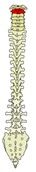
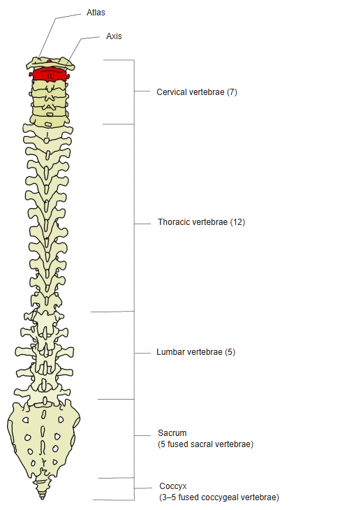
Vertebral column, axis highlighted 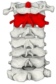
Cervical vertebrae (anterior), axis highlighted 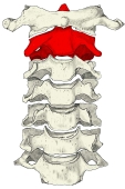
Cervical vertebrae (posterior), axis highlighted 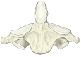
Axis (anterior) 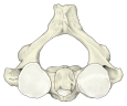
Axis (from above) 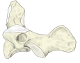
Axis (left lateral) 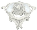
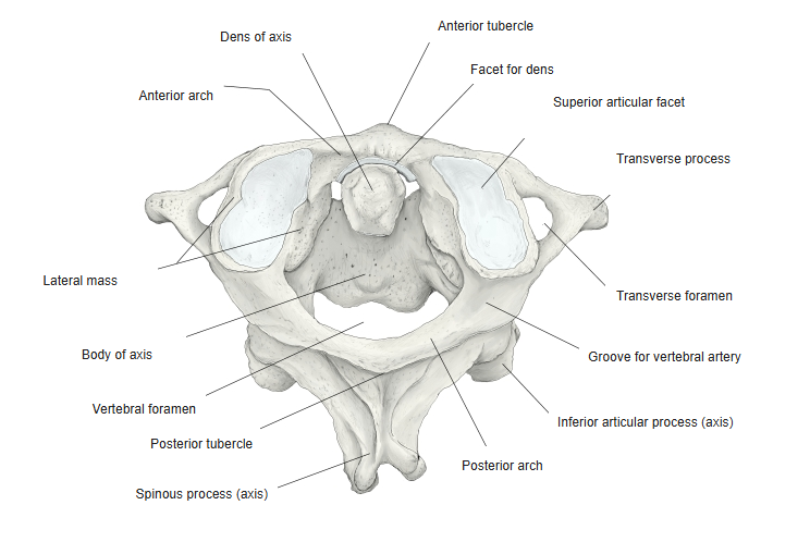
Atlas and axis (posterosuperior view) 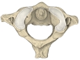
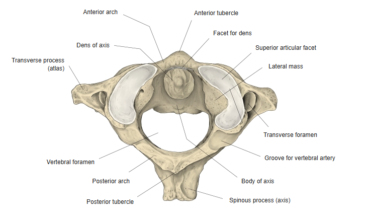
Atlas and axis (superior view) 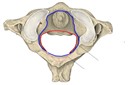
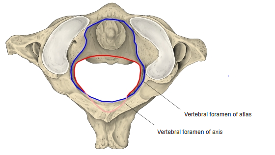
Vertebral foramina of the atlas and axis 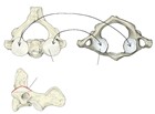
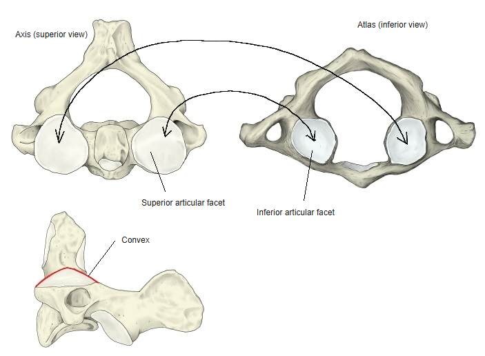
Superior articular facets of the axis and inferior articular facets of the atlas
Articulations
| # | Bone | Joint |
|---|---|---|
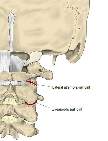
| 1 | Atlas | Lateral atlanto-axial joint (superior articular facets of axis – inferior articular facets of atlas) |
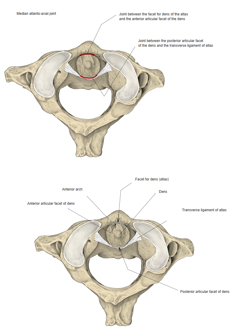
| 2 | Atlas | Median atlanto-axial joint (dens: anterior articular facet – facet for dens of atlas; posterior articular facet – transverse ligament of atlas) |
| 3 | 3rd cervical vertebra | Zygapophysial joint (inferior articular process of axis – superior articular process of C3) and intervertebral disc |
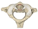
Median atlanto-axial joint 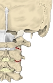
Lateral atlanto-axial and zygapophysial joints
Ligaments
The following ligaments attach to the axis. The list is intended to be comprehensive but may omit minor ligaments.
| # | Ligament | Connects |
|---|---|---|
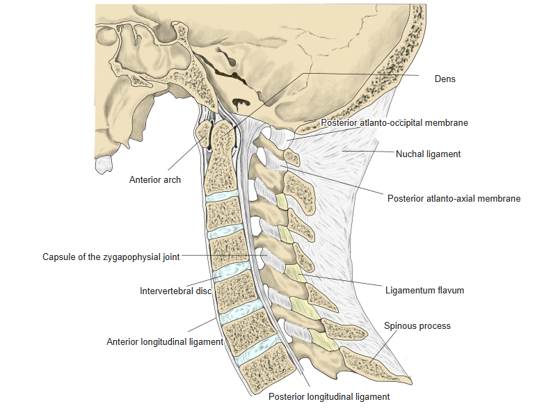
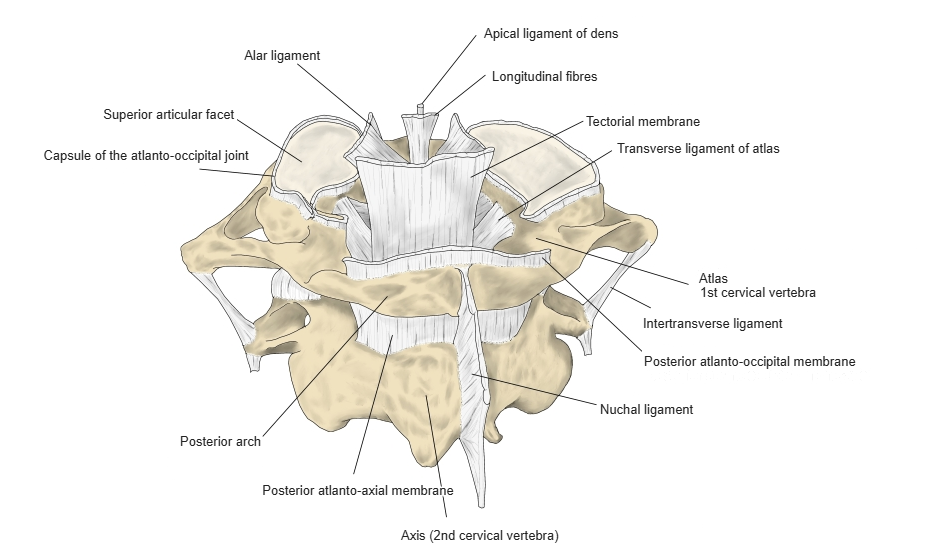
| 1 | Apical ligament of dens | Apex of the dens – front margin of the foramen magnum (occipital bone) |
| 2 | Alar ligaments | Sides of the apex of the dens – medial sides of the occipital condyles |
| 3 | Transverse ligament of atlas (with the longitudinal bands = cruciate ligament) | Holds the dens against the anterior arch of the atlas; the longitudinal bands run from the back of the body of the axis to the front margin of the foramen magnum |
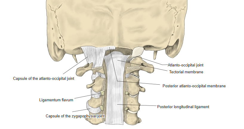
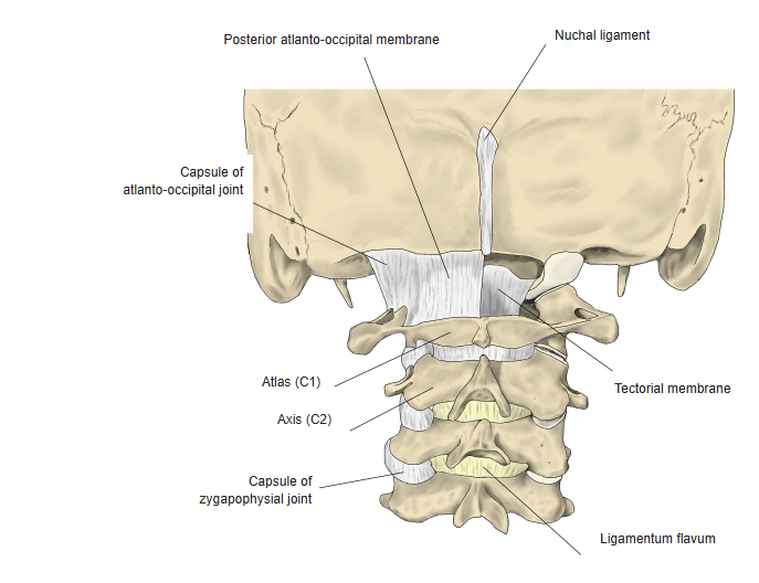
| 4 | Tectorial membrane | Back of the body of the axis – clivus (upward continuation of the posterior longitudinal ligament) |
| 5 | Anterior longitudinal ligament | Occipital bone (pharyngeal tubercle) – front of the sacrum |
| 6 | Posterior longitudinal ligament | Occipital bone (clivus) – sacral canal |
| 7 | Nuchal ligament | Occipital bone – spinous processes of the cervical vertebrae |
| 8 | Ligamenta flava | Between the laminae |
| 9 | Intertransverse ligaments | Between the transverse processes |
| 10 | Posterior atlanto-axial membrane | Arch of the axis – posterior arch of the atlas |
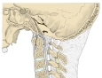
Ligaments of the cervical spine (median section) 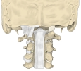
Ligaments (posterior, vertebral canal opened) 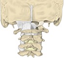
Ligaments of the craniovertebral junction (posterior) 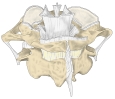
Ligaments of the atlas and axis (posterior)
Muscle attachments
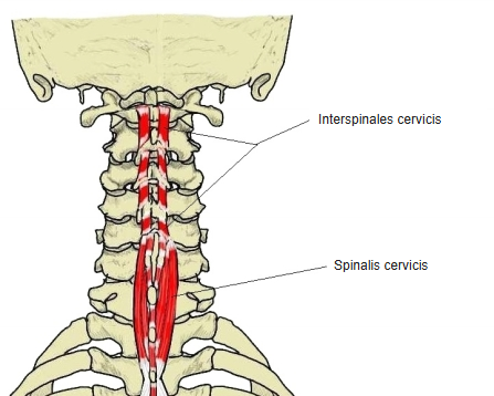
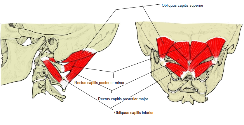
- Origin of: Rectus capitis posterior major, Obliquus capitis inferior, Interspinales cervicis
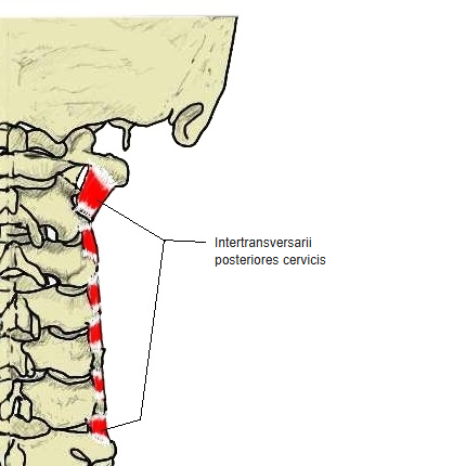
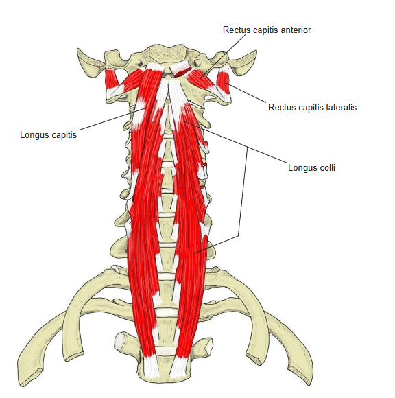
- Insertion of: Spinalis cervicis, Semispinalis cervicis, Longus colli, Intertransversarii
Rectus capitis posterior major and obliquus capitis inferior arise from the spinous process. Spinalis cervicis and semispinalis cervicis insert on the spinous process, longus colli (vertical part) on the front of the body, and intertransversarii on the transverse process. Interspinales cervicis run between the spinous processes of C2 and C3 and below.
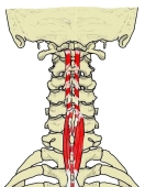
Interspinales cervicis and spinalis cervicis 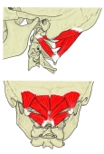
Suboccipital muscles (posterior) 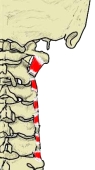
Intertransversarii posteriores cervicis 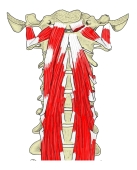
Prevertebral muscles (anterior)
Parts
- Dens (odontoid process) – the peg projecting upwards from the body.
- Apex of dens – the pointed tip of the dens; the apical ligament and the alar ligaments attach here.
- Anterior articular facet – an oval facet on the front of the dens; articulates with the facet for dens on the anterior arch of the atlas.
- Posterior articular facet – a groove-like facet on the back of the dens for the transverse ligament of the atlas.
- Body – small; its lower front edge projects downwards over the next intervertebral disc.
- Superior articular facets – large, nearly flat and roughly circular facets on either side of the dens, lying on the body and pedicles; they articulate with the inferior articular facets of the atlas.
- Inferior articular processes – lie behind the transverse processes and articulate with the 3rd cervical vertebra, as in the lower cervical vertebrae.
- Transverse process – small, with a transverse foramen; its tip corresponds to a posterior tubercle only.
- Spinous process – large, strong and bifid.
References and sources
- Further reading: Axis (anatomy), Wikipedia.