Posterior surface of ulna (Latin: facies posterior ulnae)
· It is bounded by the posterior border and the lateral border (= interosseous border).
the ulna
- Anterior surface : between the anterior border and the lateral border; faces forwards.
- Posterior surface : between the posterior border and the lateral border; faces backwards and laterally.
- Medial surface : between the anterior border and the posterior border; faces medially.
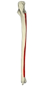 Right ulna (medial)
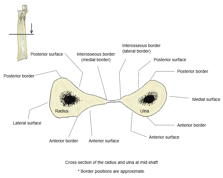 Radius and ulna (cross-section)
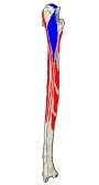 Muscle attachments (posterior)
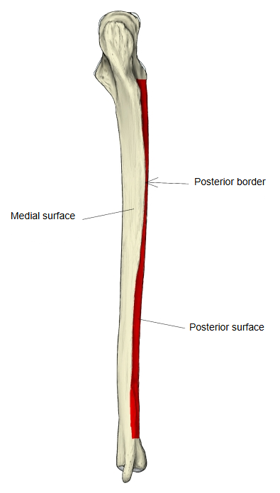
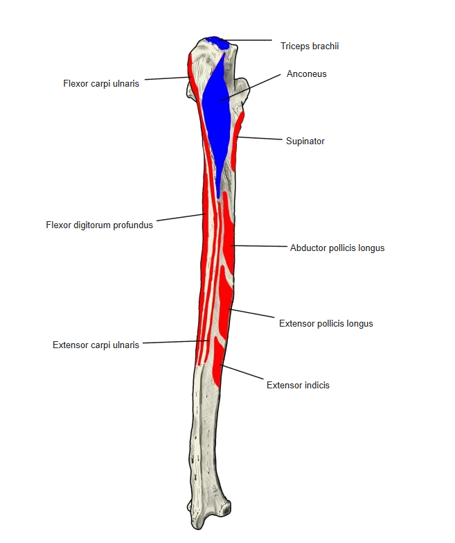
Muscles attached to the back of the ulna include anconeus, extensor carpi ulnaris, abductor pollicis longus and extensor indicis. At the lower end, on the back, a deep groove between the styloid process and the articular circumference of the head carries the tendon of extensor carpi ulnaris. On the under side, between the styloid process and the articular surface of the head, is a rough pit; here is attached the articular disc, which arises from the lower edge of the ulnar notch of the radius and separates the head of the ulna from the carpal bones.
Muscles attached to the posterior surface: anconeus, extensor carpi ulnaris, abductor pollicis longus, extensor indicis
The posterior surface is broad and hollowed above, rounded and somewhat narrower in the middle, and narrow, smooth and rounded below. At the top an oblique line runs down from the back of the radial notch to the posterior border; the triangular area above it receives the insertion of anconeus, and the upper part of the line gives attachment to supinator. Below this a vertical ridge divides the surface into a narrow, smooth medial strip covered by extensor carpi ulnaris and a wider, rougher lateral part, which gives origin, from above down, to supinator, abductor pollicis longus, extensor pollicis longus and extensor indicis.
Where it is found
Related terms
- Anterior surface of ulna
- Medial surface of ulna
- Posterior border of ulna
- Interosseous border of ulna
- Supinator crest
- Body of ulna
- Ulna
References and sources
- Further reading: Ulna, Wikipedia.
- Federative International Programme for Anatomical Terminology (FIPAT). Terminologia Anatomica, 2nd ed. (TA2), 2019.