Occipital condyle (Latin: condylus occipitalis)
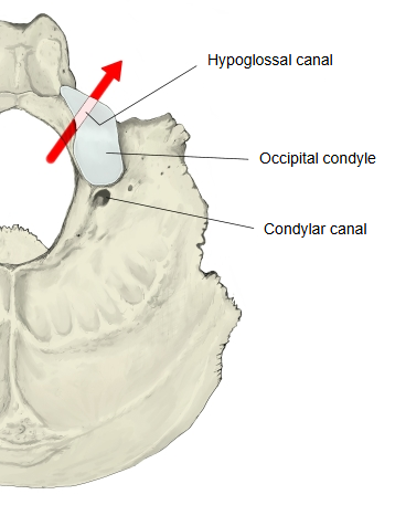
The long oval elevations on the underside of the lateral parts of the occipital bone, one on each side of the foramen magnum. They are the joint surfaces on which the skull rests on the atlas.
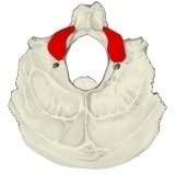 Occipital bone (inferior)
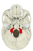 External cranial base
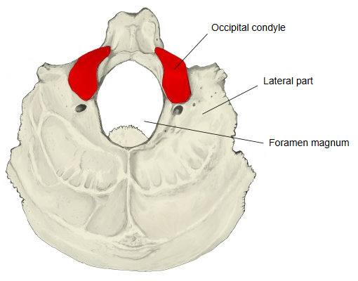
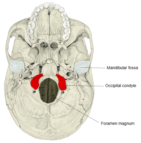
More details
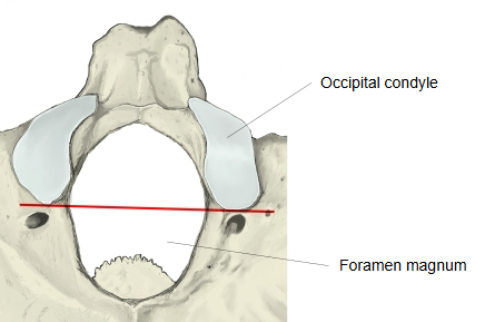
· They lie along the sides of the front half of the foramen magnum. A line joining their back ends crosses the foramen magnum at about its middle.
· Each is long and narrow, with its front end turned towards the midline. The joint surface is convex and faces downwards and laterally.
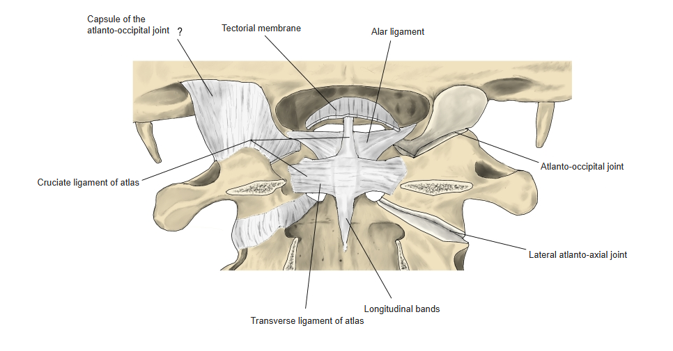
· They articulate with the superior articular facets of the atlas at the atlanto-occipital joints, which allow nodding and slight side-bending of the head.
· The hypoglossal canal runs through the bone above the base of each condyle. Behind each condyle is the condylar fossa, often pierced by a condylar canal.
· A small tubercle on the medial side of each condyle gives attachment to the alar ligaments.
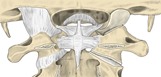 Craniovertebral joints and ligaments (posterior)
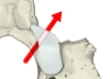 Hypoglossal canal
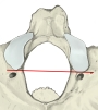 Line across the back ends of the condyles
Reference websites
[Wikipedia]
· Occipital condyles, Wikipedia.
· Atlanto-occipital joint, Wikipedia.
[Websites with illustrations]
· Occipital condyle (skull from below) (Wikimedia Commons)

· Occipital bone, outer surface, condyle marked (Gray's Anatomy, fig. 129) (Wikimedia Commons)

· The occipital bone (TeachMeAnatomy)
Where it is found
Related terms
References and sources
- Further reading: Occipital condyles, Wikipedia.
- Further reading: Atlanto-occipital joint, Wikipedia.
- Federative International Programme for Anatomical Terminology (FIPAT). Terminologia Anatomica, 2nd ed. (TA2), 2019.