Condylar canal (Latin: canalis condylaris)
Also called the posterior condylar canal
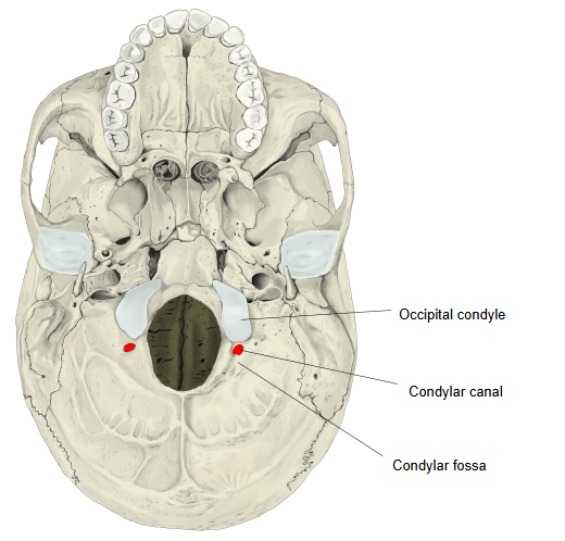
A canal of the occipital bone that opens into the condylar fossa, the hollow just behind the occipital condyle; it transmits the condylar emissary vein.
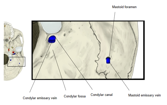
Vein: condylar emissary vein
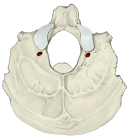 Occipital bone (inferior view)
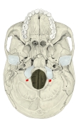 External cranial base
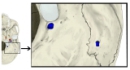 Around the condylar canal (enlarged)
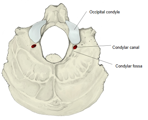
More details
The condylar canal has an internal opening into the inside of the skull and an external opening on its outer surface.
canal
- Internal opening : opens into the groove for the sigmoid sinus, close to the jugular foramen, in the posterior cranial fossa
- External opening : opens into the condylar fossa
Behind each condyle of the occipital bone there is a hollow, the condylar fossa. The posterior (external) opening of the condylar canal, a canal for a vein, lies in it, but this opening is not always present. The anterior (internal) opening of the canal lies in the groove of the sigmoid sinus.
· The condylar emissary vein links the sigmoid sinus with the veins outside the skull (the suboccipital venous plexus and the vertebral venous plexuses); a small meningeal branch of the occipital artery may run with it.
· The canal is often absent on one or both sides, or ends blindly without passing through the bone. Sources differ on how often it is present; figures from about half to most skulls are quoted, and it is frequently present on one side only.
· It should not be confused with the hypoglossal canal, which lies above and in front of the condyle and is sometimes called the anterior condylar canal.
 Foramina of the external cranial base
 Foramina of the internal cranial base
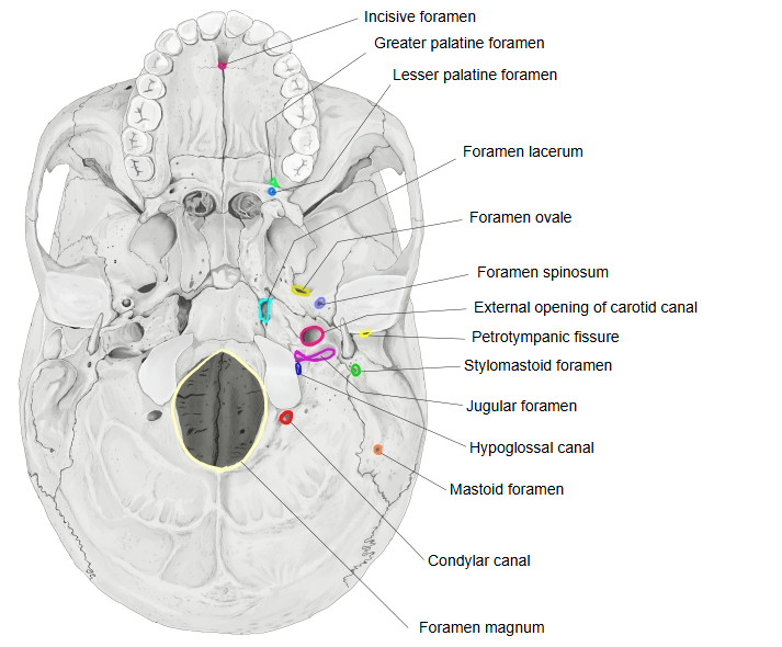
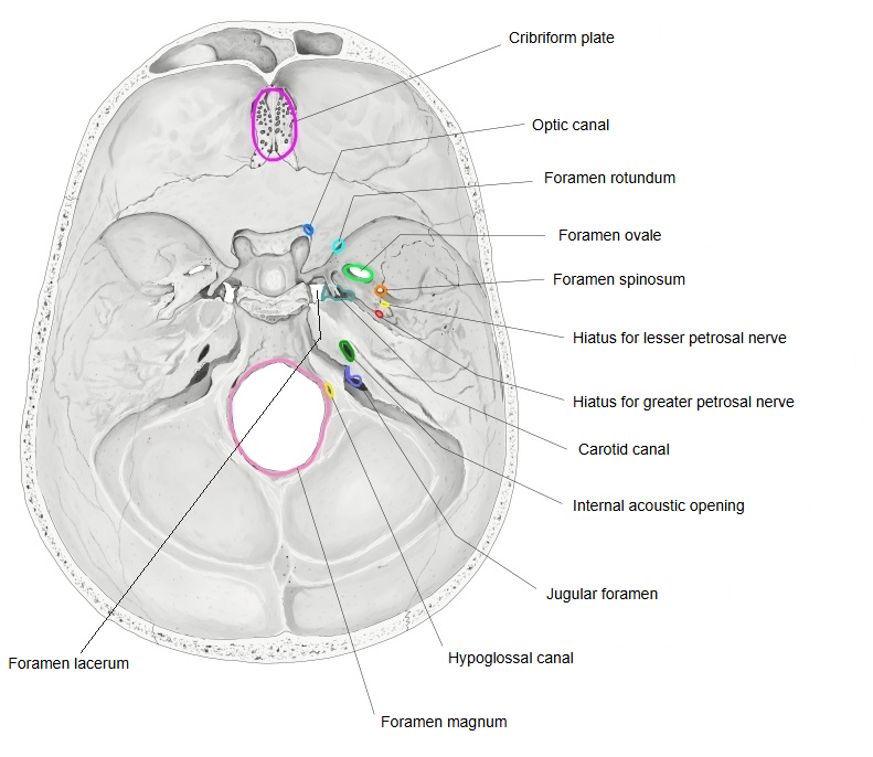
Reference websites
[Wikipedia]
· Condylar canal, Wikipedia.
[Websites with illustrations]
· Condylar canal (Wikimedia Commons)

· Occipital bone, outer surface (Gray's Anatomy, fig. 129) (Wikimedia Commons)

· The occipital bone (TeachMeAnatomy)
Where it is found
Related terms
- Occipital condyle
- Condylar emissary vein
- Sigmoid sinus
- Hypoglossal canal
- Jugular foramen
- Foramen magnum
- Emissary veins
References and sources
- Further reading: Condylar canal, Wikipedia.
- Federative International Programme for Anatomical Terminology (FIPAT). Terminologia Anatomica, 2nd ed. (TA2), 2019.