Orbit (Latin: orbita)
Also called Eye socket
- Overview
- Bones forming the orbit (four walls)
- Orbital margins
- Openings
- Muscles
- Ligaments
- Nerves
- Vessels (arteries and veins)
- Other
- Reference websites
- (Wikipedia, websites with illustrations)
- Where it is found
- Related terms
Overview
· In an adult the orbit holds about 30 mL, of which the eyeball takes up about 6.5-7 mL.
Japanese anatomy textbooks describe its shape as follows.
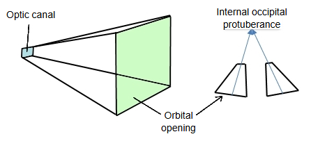
Each orbit is shaped like a four-sided pyramid. Its base faces forwards and laterally and is the orbital opening on the face; its apex points backwards and medially to the optic canal. The long axes of the two orbits therefore diverge forwards; traced backwards, they meet far behind the orbits, roughly at the level of the internal occipital protuberance.
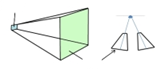 Shape and axes of the orbits
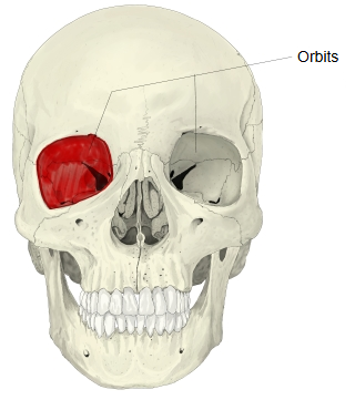
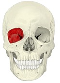 Orbits (skull, anterior)
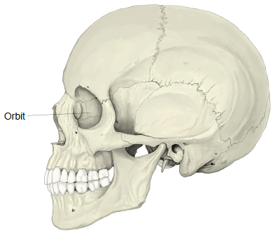
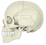 Orbit (skull, lateral)
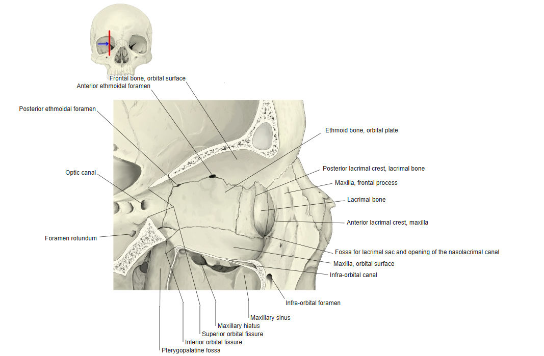
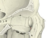 Right orbit, section (lateral view)
The main contents of the orbit are listed below.
of the
orbit
- Eyeball : front-to-back diameter about 24 mm. Main parts: cornea, anterior chamber, lens, vitreous body.
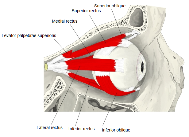
- Muscles : intrinsic muscles (such as the ciliary muscle), extra-ocular muscles (such as the superior rectus) and others (such as the levator palpebrae superioris).
- Vessels : arteries: ophthalmic artery, supratrochlear artery, lacrimal artery and others; veins: superior ophthalmic vein, lacrimal vein and others.
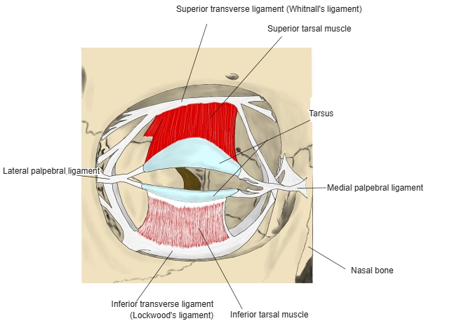
- Ligaments : Lockwood's ligament, Whitnall's ligament and others.
- Nerves : optic nerve, nasociliary nerve, lacrimal nerve and others, with the ciliary ganglion.
- Others : the lacrimal apparatus (lacrimal gland, lacrimal sac and others) and fat.
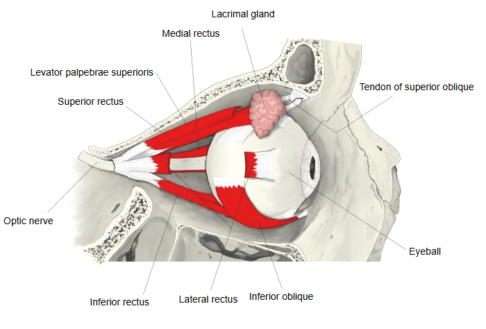
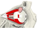 Extra-ocular muscles (lateral)
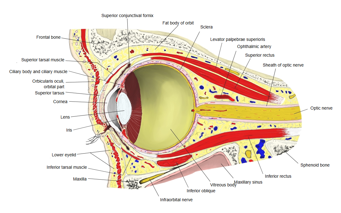
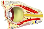 Orbit, sagittal section
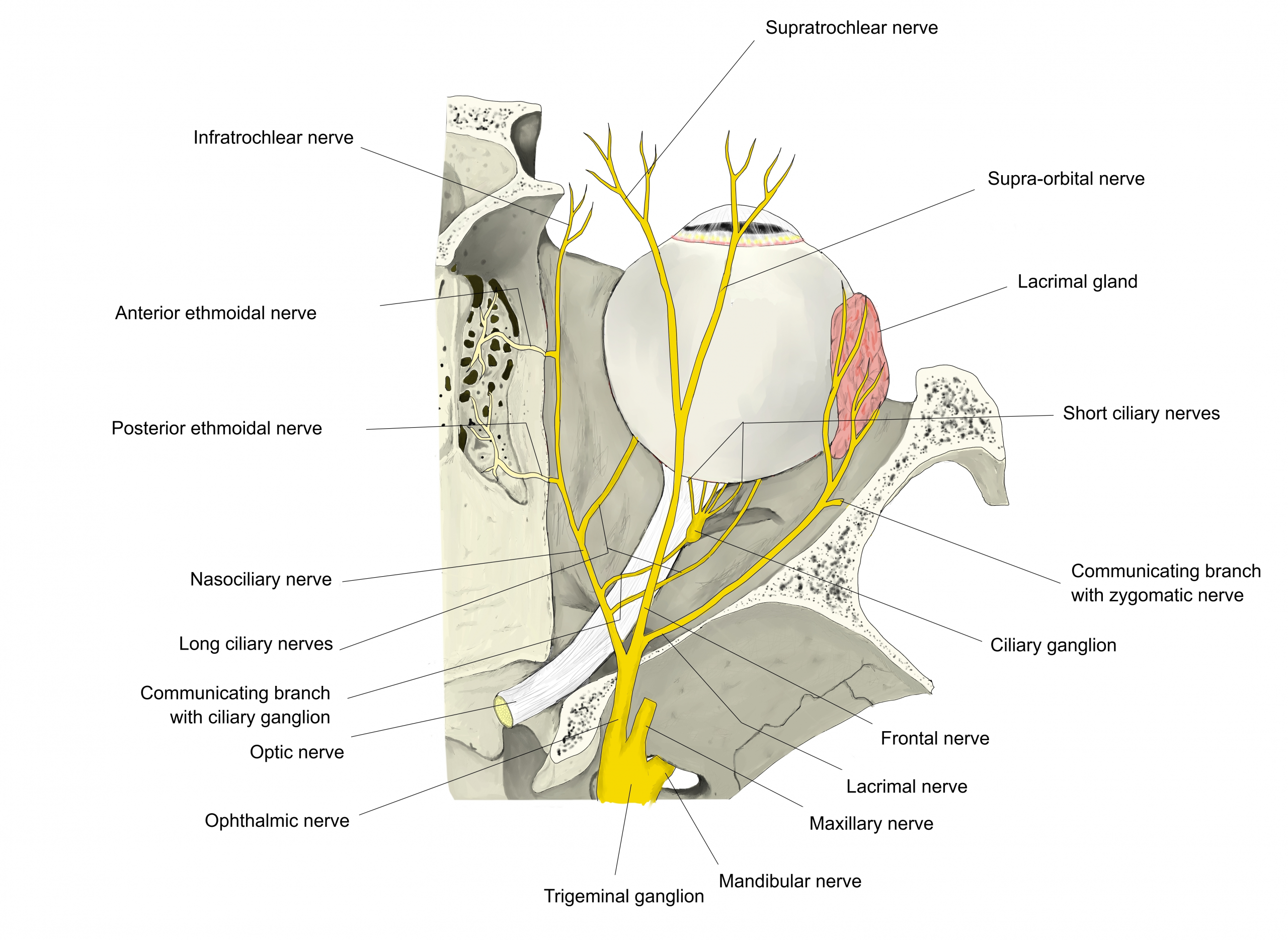
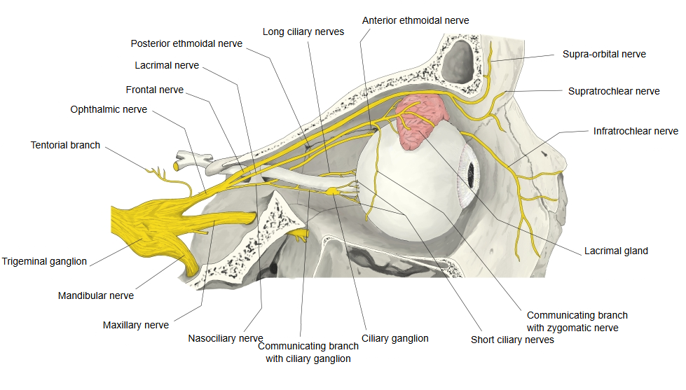
 Nerves of the orbit (from above)
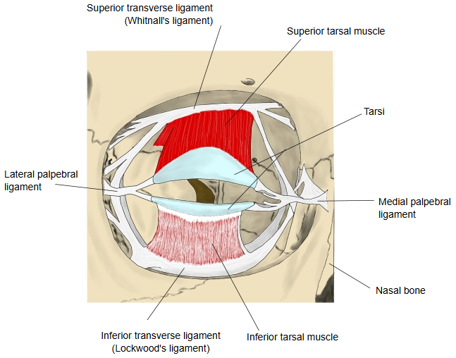
 Ligaments of the eyelids
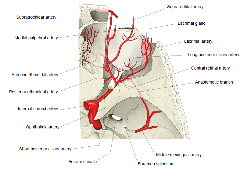
 Ophthalmic artery (from above)
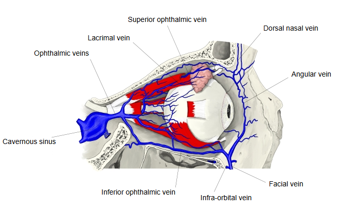
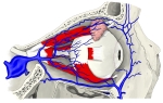 Ophthalmic veins (lateral)
Bones forming the orbit (four walls)
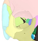 Right orbit (bones colour-coded)
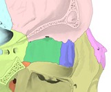 Medial wall (bones colour-coded)
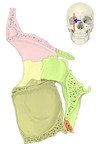 Lateral wall, section (bones colour-coded)
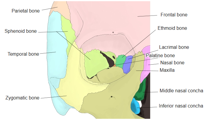
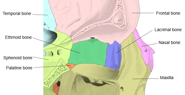
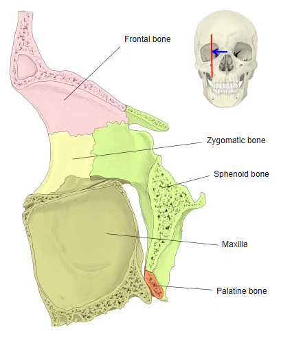
[The four walls]
The orbit can be divided into four walls: the roof (superior wall), the floor (inferior wall), the medial wall and the lateral wall.
| No | Wall | Bones | Features | Figure |
|---|---|---|---|---|
| 1 | Roof (superior wall) | · Frontal bone (orbital part) · Sphenoid bone (lesser wing) | · Optic canal (at the apex) · Trochlear fovea (medially) · Fossa for the lacrimal gland (laterally) | 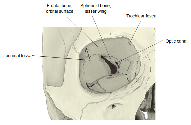 |
| 2 | Floor (inferior wall) | · Maxilla (orbital surface) · Zygomatic bone (orbital surface) · Palatine bone (orbital process) | · Infra-orbital groove · Infra-orbital canal | 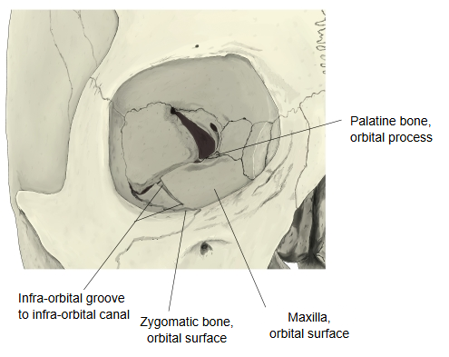 |
| 3 | Medial wall · the thinnest wall · often bulges slightly | · Ethmoid bone (orbital plate, also called the lamina papyracea) · Sphenoid bone (body) · Lacrimal bone · Maxilla (frontal process) | · Anterior and posterior lacrimal crests · Fossa for the lacrimal sac · Anterior and posterior ethmoidal foramina The thin ethmoidal part breaks easily in injuries of the orbit. | 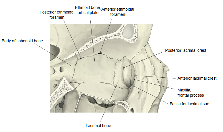 |
| 4 | Lateral wall · the thickest wall | · Sphenoid bone (orbital surface of the greater wing) · Zygomatic bone (orbital surface) | · Superior orbital fissure · Inferior orbital fissure · Zygomatico-orbital foramen | 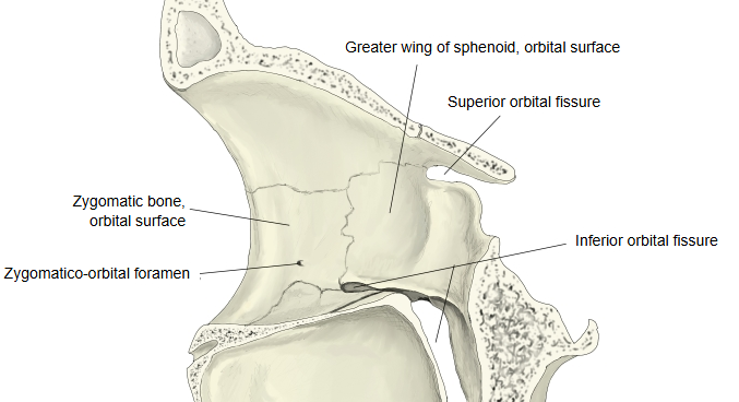 |
Orbital margins
| 1 | Supra-orbital margin | Frontal bone; it carries the supra-orbital notch (or foramen) and the frontal notch (or foramen). |
| 2 | Infra-orbital margin | Maxilla and zygomatic bone. |
| 3 | Medial margin | Frontal bone and maxilla (frontal process). |
| 4 | Lateral margin | Zygomatic bone and frontal bone. |
Openings
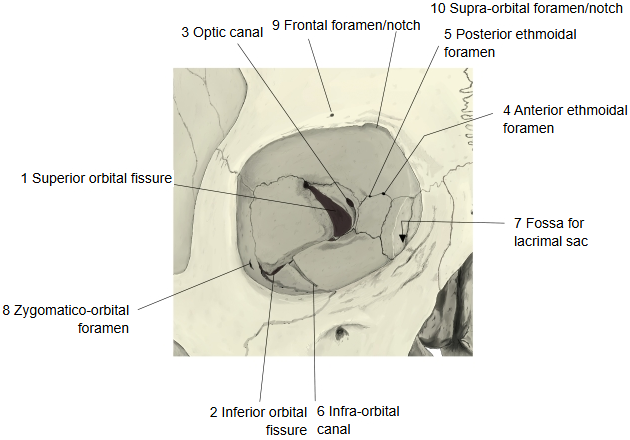
Through the following foramina and fissures the orbit communicates with other parts of the skull, and nerves and vessels pass through them.
Muscles
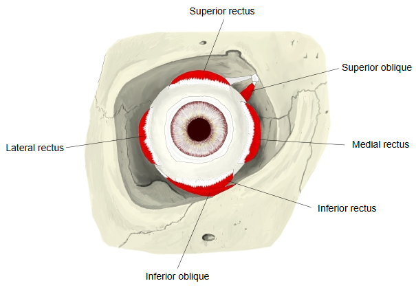
Here the muscles of the orbit are divided into three groups.
The muscles deep to the orbicularis oculi are counted as muscles of the orbit; the list may not be complete.
| 1 | Intrinsic muscles (inside the eyeball) | 1 | Ciliary muscle | 3 | Dilator pupillae (muscle of the iris) |
| 2 | Sphincter pupillae (muscle of the iris) | ||||
| 2 | Extra-ocular muscles (move the eyeball) | 1 | Lateral rectus | 4 | Inferior rectus |
| 2 | Medial rectus | 5 | Inferior oblique | ||
| 3 | Superior rectus | 6 | Superior oblique | ||
| 3 | Others | 1 | Levator palpebrae superioris (raises the upper eyelid) | 4 | Orbitalis (thin smooth muscle bridging the inferior orbital fissure) |
| 2 | Superior tarsal muscle (smooth muscle) | 5 | Tensor trochleae (an occasional slip) | ||
| 3 | Inferior tarsal muscle (smooth muscle) | 6 | Muscle of Riolan (ciliary part of the orbicularis oculi at the lid margin) |
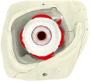 Extra-ocular muscles (anterior)
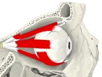 Extra-ocular muscles (lateral)
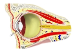 Eyeball in the orbit (sagittal section)
Ligaments
The ligaments of the orbit lie around the eyelids and the front of the orbit; the back of the orbit seems to have no true ligaments.
of the
orbit
- Whitnall's ligament : a band above the eyeball, formed by the thickened sheath of the levator palpebrae superioris. = superior transverse ligament
- Lockwood's ligament : a hammock-like band below the eyeball that supports it (suspensory ligament of the eyeball). Sources differ on whether it is the same as the inferior transverse ligament.
- Medial palpebral ligament : a short band (about 4 mm long, 2 mm wide) at the inner corner of the eye that joins the upper and lower tarsi to the medial orbital margin.
- Lateral palpebral ligament : a band at the outer corner of the eye that ties the tarsi to the lateral orbital tubercle.
Nerves
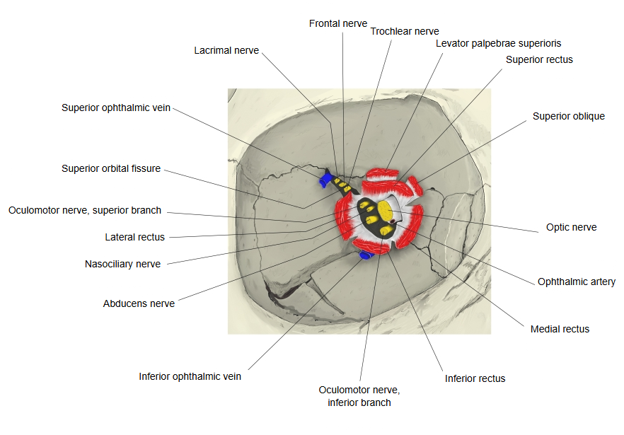
The nerves of the orbit, including several cranial nerves, are listed below; the list may not be complete.
| Nerve | Route into the orbit | Notes | |
|---|---|---|---|
| 1 | Oculomotor nerve (III) | Superior orbital fissure | To the extra-ocular muscles and levator palpebrae superioris |
| 2 | Trochlear nerve (IV) | Superior orbital fissure | To the superior oblique |
| 3 | Ophthalmic nerve (V1, first division of the trigeminal nerve) | Superior orbital fissure | Divides into the lacrimal, frontal (supra-orbital and supratrochlear) and nasociliary nerves; the nasociliary nerve gives the long ciliary nerves, the anterior and posterior ethmoidal nerves and the infratrochlear nerve. |
| 4 | Abducens nerve (VI) | Superior orbital fissure | To the lateral rectus |
| 5 | Infra-orbital nerve (from V2) | Inferior orbital fissure → infra-orbital canal | |
| 6 | Zygomatic nerve (from V2) | Inferior orbital fissure → zygomatico-orbital foramen | |
| 7 | Optic nerve (II) | Optic canal | The ciliary ganglion lies lateral to it and sends the short ciliary nerves to the eyeball. |
| 8 | Anterior ethmoidal nerve | Anterior ethmoidal foramen | |
| 9 | Posterior ethmoidal nerve | Posterior ethmoidal foramen | |
| 10 | Supra-orbital nerve, medial branch | Frontal notch (or foramen) | |
| 11 | Supra-orbital nerve, lateral branch | Supra-orbital notch (or foramen) |
- Nerves of the orbit (from above)
 Ophthalmic nerve (lateral)
 Orbital apex (anterior)
- Eyeball in the orbit (sagittal section)
Vessels (arteries and veins)
The vessels found in the orbit are listed below; the lists may not be complete.
[Arteries]
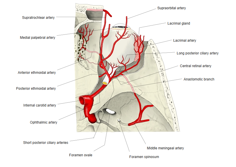
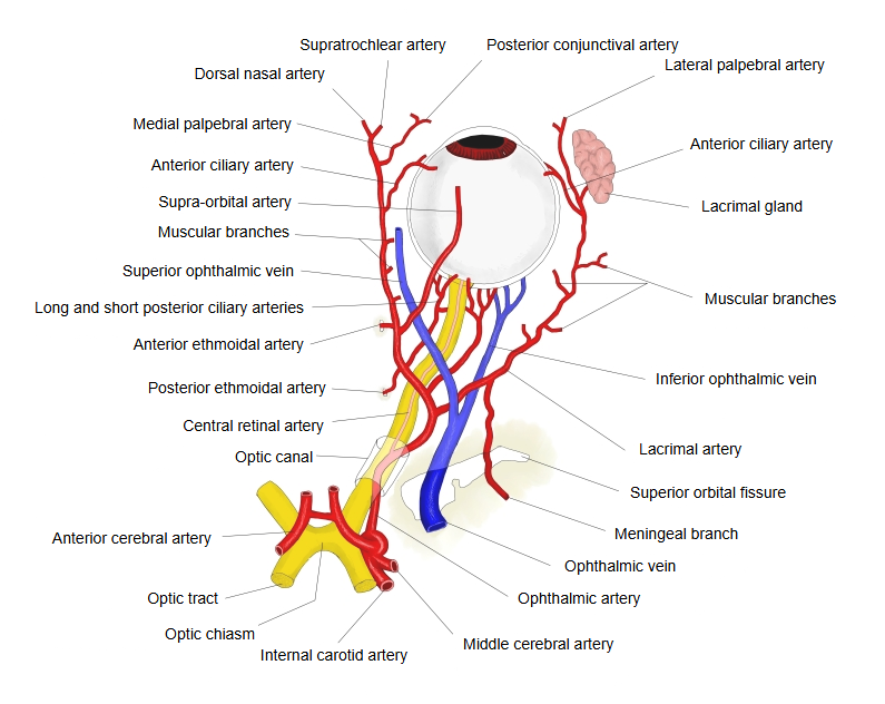
Most arteries of the orbit are branches of the ophthalmic artery, which enters through the optic canal.
| Artery | Notes | |
|---|---|---|
| 1 | Ophthalmic artery | The first large branch of the internal carotid artery; the source of most arteries of the orbit. |
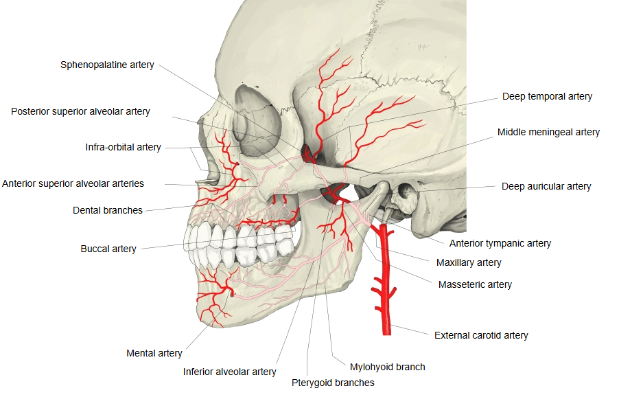
| 2 | Infra-orbital artery | A branch of the maxillary artery (external carotid system); runs in the floor of the orbit with the infra-orbital nerve and comes out through the infra-orbital foramen. |
| 3 | Supra-orbital artery | Runs forwards under the roof of the orbit and leaves it through the supra-orbital notch. |
| 4 | Anterior ethmoidal artery | Goes with the anterior ethmoidal nerve through the anterior ethmoidal foramen towards the nasal cavity. |
| 5 | Lacrimal artery | Leaves the ophthalmic artery lateral to the optic nerve and goes to the lacrimal gland. Branch: lateral palpebral arteries, to the lateral halves of the upper and lower eyelids at the lateral angle of the eye. |
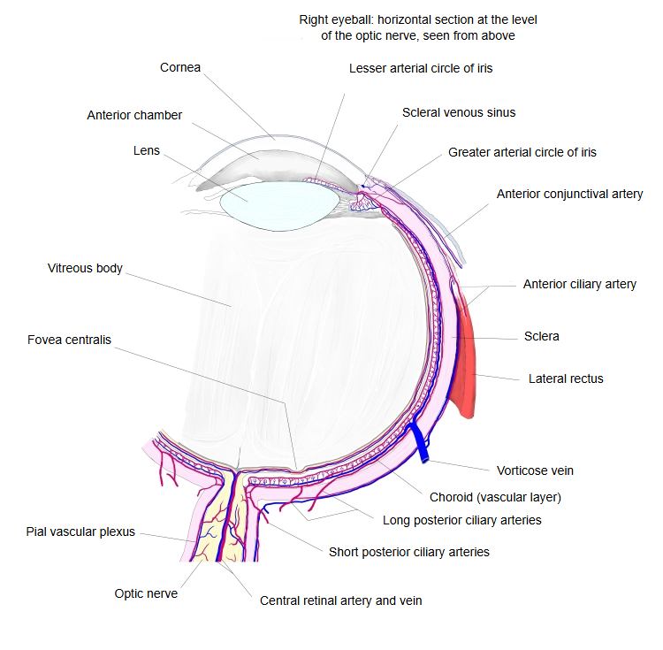
| 6 | Short posterior ciliary arteries | Pierce the sclera at the back of the eyeball and supply the choroid. |
| 7 | Long posterior ciliary arteries | A medial and a lateral one; they run forwards to the ciliary body and iris and form the arterial circle of the iris. |
| 8 | Medial palpebral arteries | An upper and a lower branch; they join the lateral palpebral arteries to form the upper and lower palpebral arches. |
| 9 | Central retinal artery | Enters the optic nerve and runs in it to the retina. |
| 10 | Anterior ciliary arteries | Come from the muscular branches of the ophthalmic artery and enter the wall of the eyeball just behind the corneal limbus. Branches: episcleral arteries, anterior and posterior conjunctival arteries. |
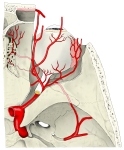 Ophthalmic artery
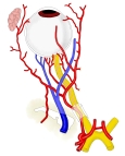 Branches of the ophthalmic artery
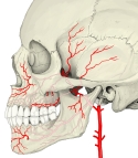 Maxillary artery
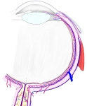 Vessels of the eyeball
[Veins]
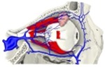
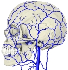
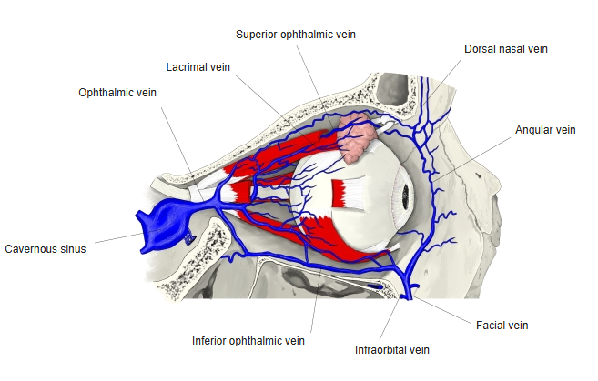
The ophthalmic veins drain mainly into the cavernous sinus and also connect with the facial (angular) vein, so infections of the face can spread into the skull.
| Vein | Notes | |
|---|---|---|
| 1 | Ophthalmic veins | A collective name for the superior and inferior ophthalmic veins, or the trunk formed when they join. |
| 2 | Superior ophthalmic vein | Collects the venous blood from the area supplied by the ophthalmic artery; leaves through the superior orbital fissure. |
| 3 | Inferior ophthalmic vein | Drains the floor of the orbit and joins the superior ophthalmic vein (or the pterygoid plexus). |
| 4 | Lacrimal vein | A tributary of the superior ophthalmic vein; collects blood from the lacrimal gland. |
| 5 | Vorticose veins | Tributaries of the ophthalmic veins; formed by the veins of the choroid and ciliary body. |
| 6 | Anterior ciliary veins | Tributaries of the superior ophthalmic vein; collect blood from the ciliary body. |
| 7 | Episcleral veins | Fine veins on the surface of the sclera; tributaries of the superior ophthalmic vein. |
| 8 | Central retinal vein | Formed by the veins of the retina; drains into the superior ophthalmic vein or directly into the cavernous sinus. |
 Superior ophthalmic vein
- Branches of the ophthalmic artery
 Veins of the head
- Vessels of the eyeball
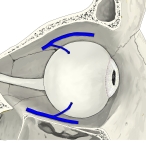 Vorticose veins
Other
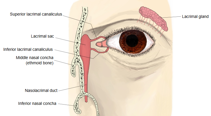
[Lacrimal apparatus]
The orbit contains the organs that make tears and those that carry them away: the lacrimal gland makes the tears, and the lacrimal canaliculi, the lacrimal sac and the nasolacrimal duct carry them to the nose.
[Fat]
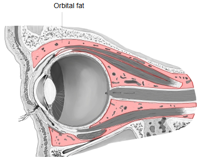
The spaces between the eyeball, the muscles, the vessels and the nerves in the orbit are filled with fat (adipose tissue), which holds each of them in place.
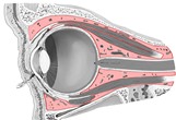 Orbital fat
- Eyeball in the orbit (sagittal section)
Reference websites
[Wikipedia]
· Orbit (anatomy), Wikipedia.
· Ophthalmic artery, Wikipedia.
· Extraocular muscles, Wikipedia.
[Websites with illustrations]
· The orbit (Kenhub)
· The bony orbit (TeachMeAnatomy)
· Orbit (Radiopaedia)
Where it is found
Related terms
- Superior orbital fissure
- Supra-orbital foramen
- Infraorbital foramen
- Orbital plate
- Fossa for lacrimal sac
- Posterior lacrimal crest
- Zygomatico-orbital foramen
References and sources
- Further reading: Orbit (anatomy), Wikipedia.
- Further reading: Ophthalmic artery, Wikipedia.
- Further reading: Extraocular muscles, Wikipedia.
- Federative International Programme for Anatomical Terminology (FIPAT). Terminologia Anatomica, 2nd ed. (TA2), 2019.