Latin: Arcus aortae
Overview
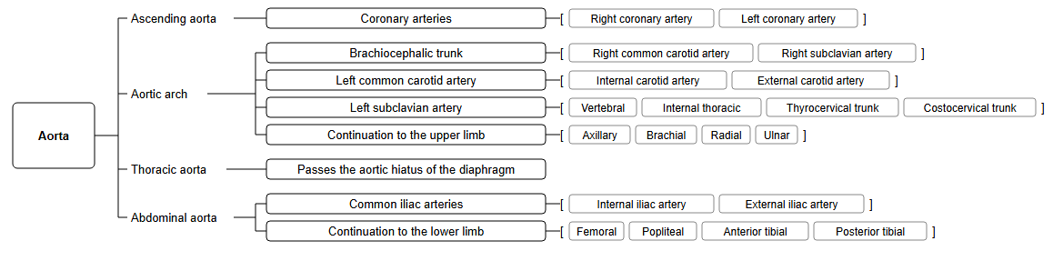
- The curved part of the aorta between the ascending aorta and the thoracic (descending) aorta.
- Gives off three large branches to the head, neck and upper limbs: the brachiocephalic trunk, the left common carotid artery and the left subclavian artery.
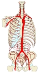
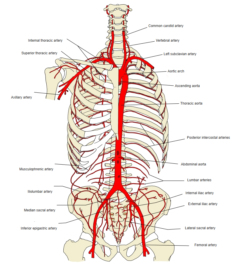
Aorta and its branches in the trunk (front view) 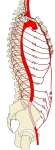
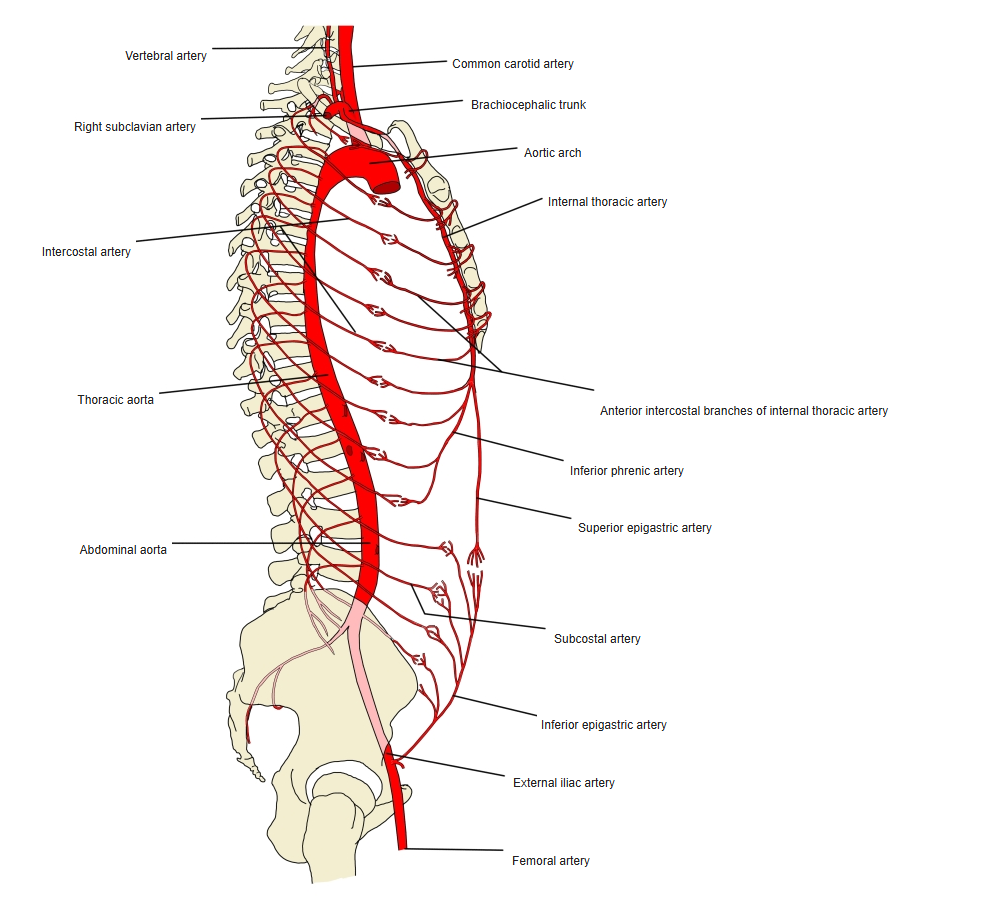
Aorta and its branches in the trunk (side view) 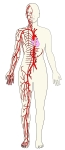
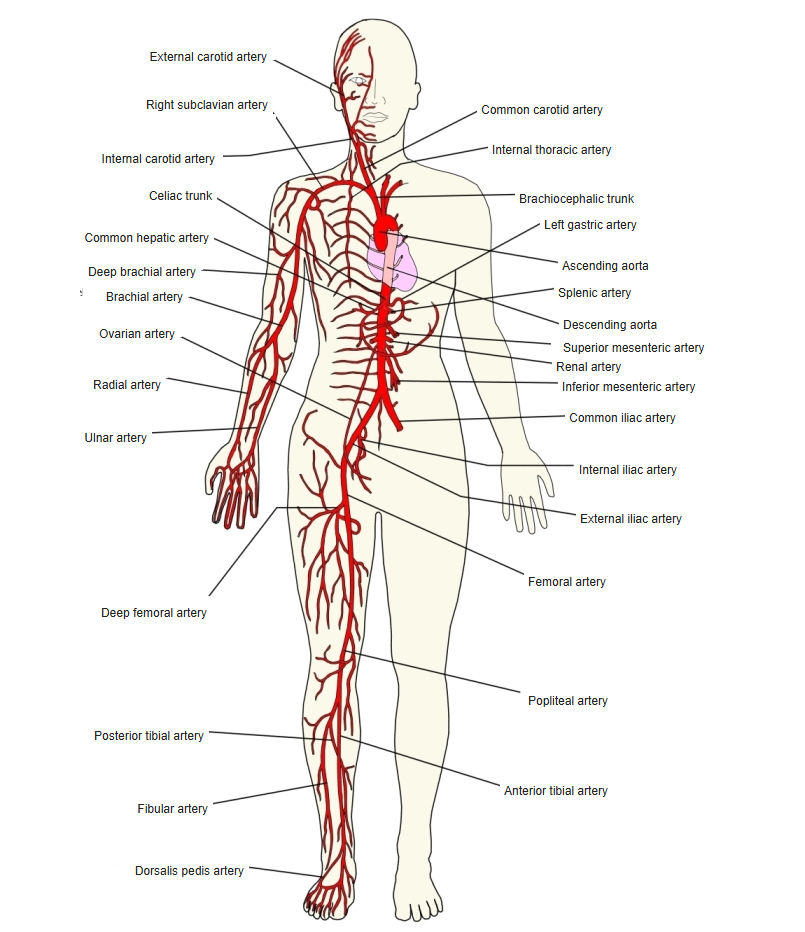
Main arteries of the body 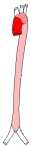
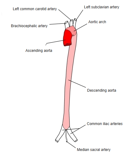
Parts of the aorta
Course
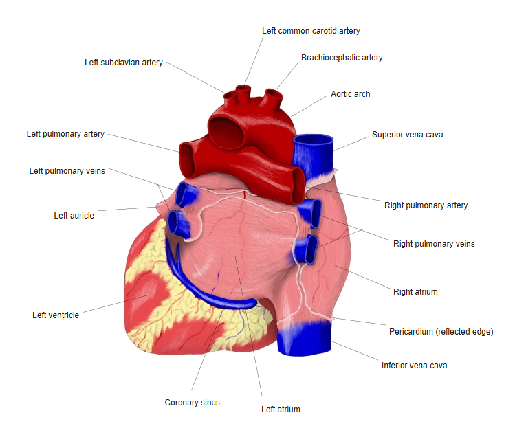
- Begins at the level of the 2nd right sternocostal joint and arches backward and to the left, over the root of the left lung.
- Ends at the left side of the 4th thoracic vertebra, where it continues as the thoracic aorta.
- The ligamentum arteriosum (remnant of the fetal ductus arteriosus) connects its lower surface to the left pulmonary artery.
Aortic arch and the heart (back view) 
Aortic arch and the heart (front view)
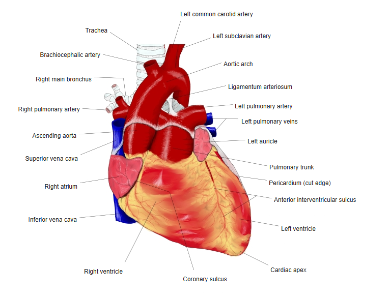
Branches
| Branch | Further course |
|---|---|
| Brachiocephalic trunk | Divides into the right subclavian and right common carotid arteries |
| Left common carotid artery | Ascends in the neck and divides into the internal and external carotid arteries |
| Left subclavian artery | Supplies the left upper limb, the neck and the brain (vertebral artery) |
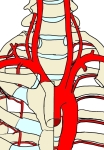
Branches of the aortic arch
Relations
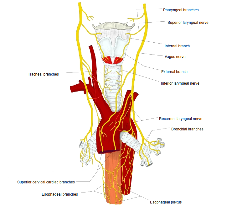
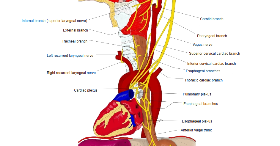
- The left recurrent laryngeal nerve hooks under the aortic arch behind the ligamentum arteriosum.
- The trachea and esophagus lie to its right; the vagus and phrenic nerves cross its left side.
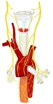
Vagus nerves around the aortic arch 
Aortic arch and nearby nerves (side view)
References and sources
- Further reading: Aortic arch, Wikipedia.