Latin: Vena azygos
Overview
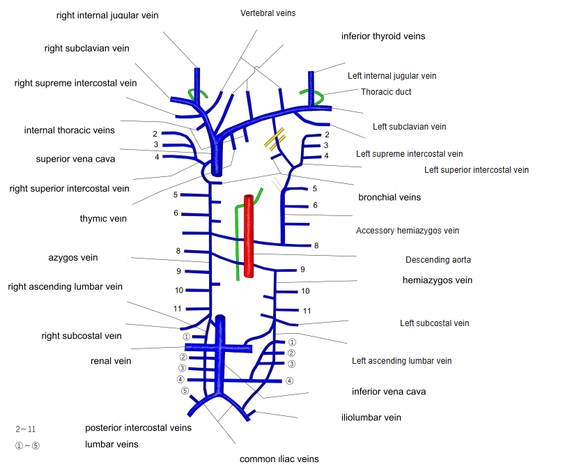
- An unpaired vein on the right side of the vertebral bodies in the posterior mediastinum.
- Usually begins from the right ascending lumbar vein and the right subcostal vein (sometimes from the inferior vena cava).
- Enters the thorax through the aortic hiatus (or the right crus of the diaphragm).
- Arches forward over the root of the right lung at about T4 and ends in the superior vena cava.
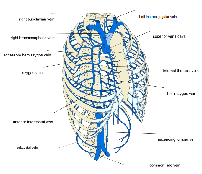
Azygos system (front view) 
Superior vena cava and its tributaries
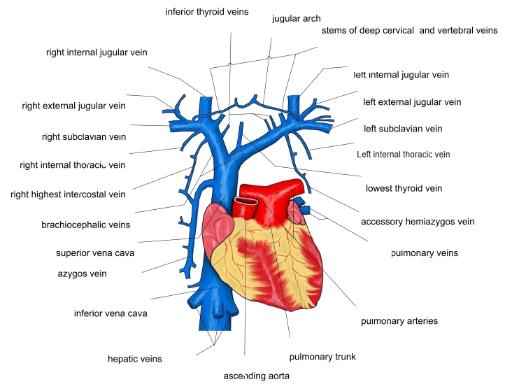
Tributaries
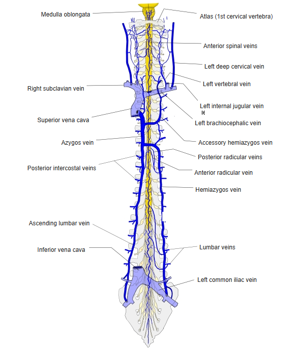
- Right posterior intercostal veins (the upper ones through the right superior intercostal vein).
- Hemiazygos and accessory hemiazygos veins.
- Esophageal, bronchial, mediastinal and pericardial veins.
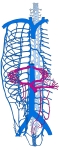
Veins of the posterior thoracic wall 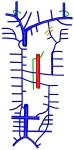
Veins of the thorax and their drainage 
Veins of the vertebral column and posterior trunk wall
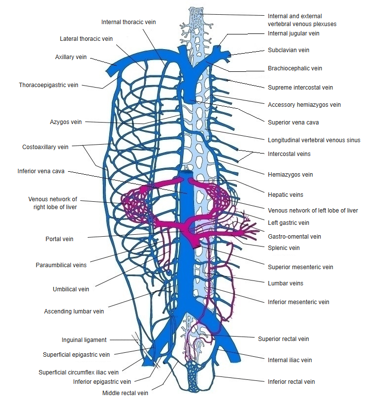
Relations
- The thoracic duct and the descending aorta lie on its left.
- The esophagus lies in front of it.
Clinical notes
- Enlarges when the superior or inferior vena cava is obstructed, carrying blood between them.
References and sources
- Further reading: Azygos vein, Wikipedia.