Thoracic duct (Latin: ductus thoracicus)
- Overview
- Course
- Reference websites
- (Wikipedia, websites with illustrations)
- Where it is found
- Related terms
Overview
· Length: about 38–45 cm in adults. Width: usually about 2–5 mm. It has many valves and often looks beaded.
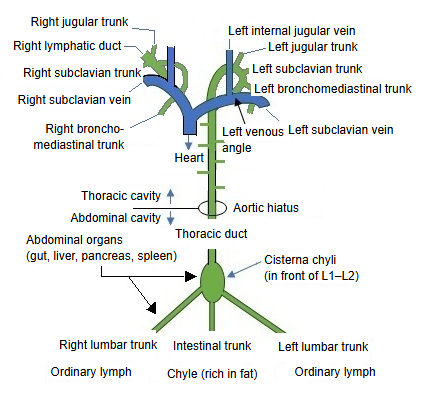
The path of chyle
Chyle is the milky, fat-rich lymph of the intestine; the thoracic duct carries it together with ordinary lymph.
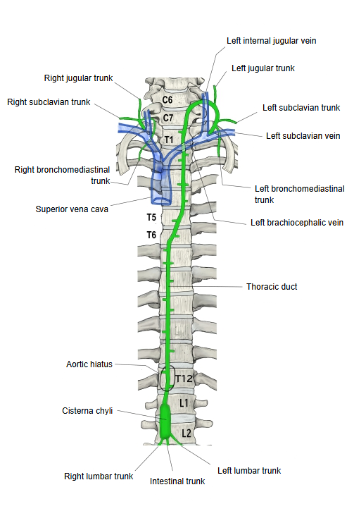
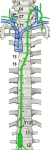 Thoracic duct on the spine
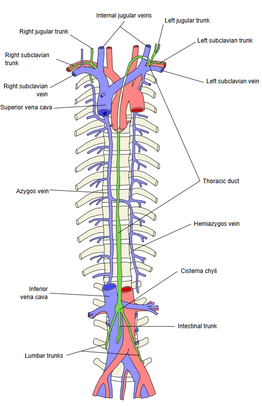
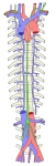 Thoracic duct and the azygos system
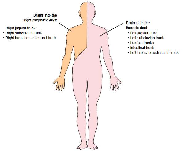
 Lymph vessels of the trunk
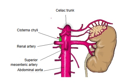
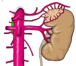 Cisterna chyli and the abdominal aorta
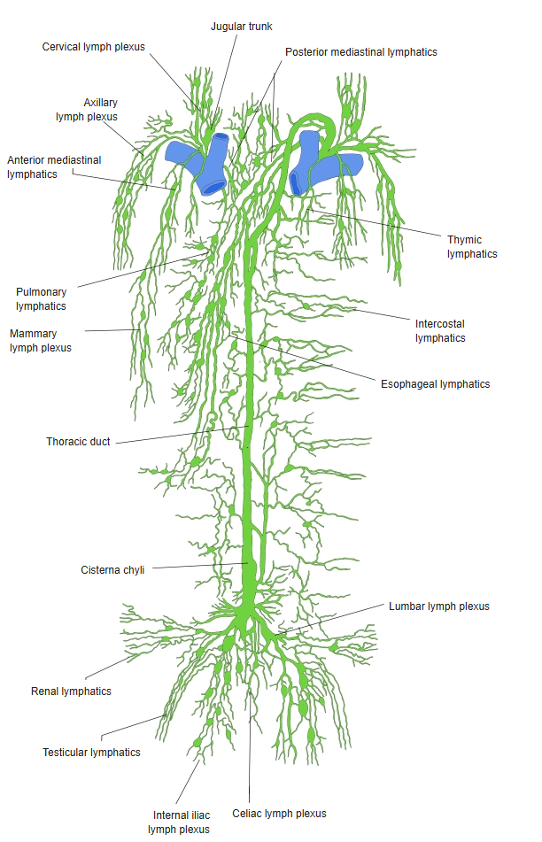
From lymph capillaries to the veins
The thoracic duct is sometimes counted as a kind of lymphatic trunk. It begins with a dilatation, the cisterna chyli, and carries about three quarters of the body's lymph to the veins.
Course
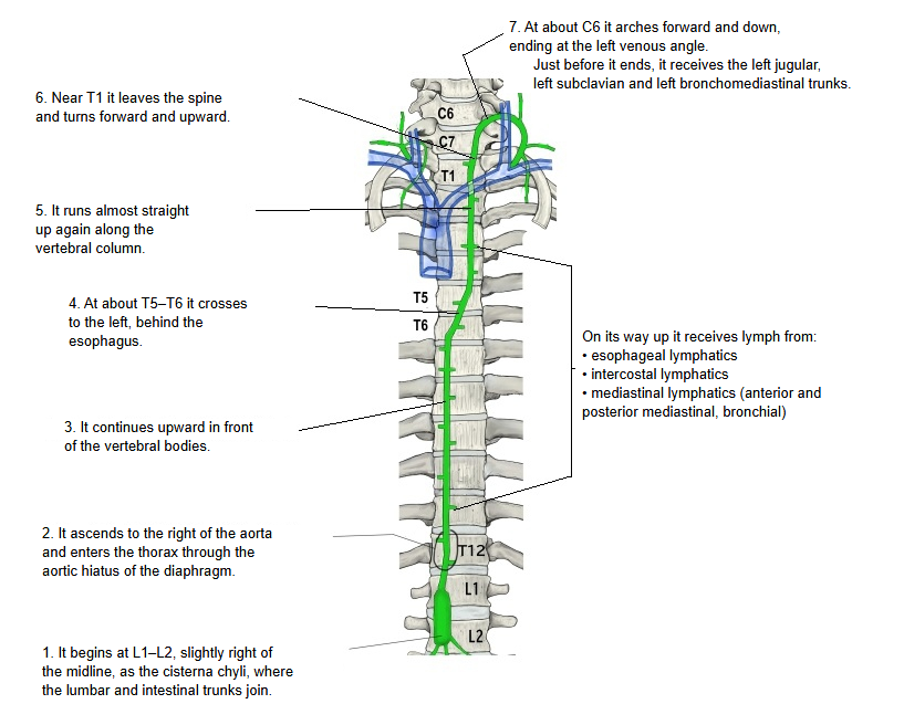
1. 1. It begins in front of the first or second lumbar vertebra (slightly right of the midline) as a dilatation, the cisterna chyli, where the lymph of the right and left lumbar trunks and the intestinal trunk comes together.
2. 2. It rises in front of the vertebral column, on the right side of the aorta, and passes with the aorta through the aortic hiatus of the diaphragm (about T12) into the thoracic cavity.
3. 3. It continues upward in front of the vertebral bodies, in the posterior mediastinum between the thoracic aorta and the azygos vein, behind the esophagus.
4. 4. At about the fifth or sixth thoracic vertebra it turns to the left, crossing behind the esophagus, and comes to lie on the left side of the esophagus.
5. 5. It then runs almost straight up again along the vertebral column into the superior mediastinum.
On its way up in front of the vertebral column, the thoracic duct receives lymph from these lymph vessels:
· Esophageal lymph vessels · intercostal lymph vessels · mediastinal lymph vessels (anterior and posterior mediastinal lymph vessels, right and left bronchial lymph vessels)
6. 6. At about the top of the first thoracic vertebra (the root of the neck) it leaves the vertebral column and turns forward, upward and laterally, behind the carotid sheath.
7. 7. When its upper end reaches about the level of C6–C7, it arches downward and opens into the veins at the left venous angle, the junction of the left internal jugular vein and the left subclavian vein. From there the lymph flows through the left brachiocephalic vein and the superior vena cava back to the heart.
Just before it opens into the vein, it receives the lymph of the left jugular trunk, the left subclavian trunk and the left bronchomediastinal trunk; any of these may instead open separately into the veins. The end of the duct is often doubled or forms a plexus (see lymphatic plexus).
Reference websites
[Wikipedia]
· Thoracic duct, Wikipedia.
· Cisterna chyli, Wikipedia.
[Websites with illustrations]
· Thoracic duct in the posterior thorax (slide) (SlideShare)

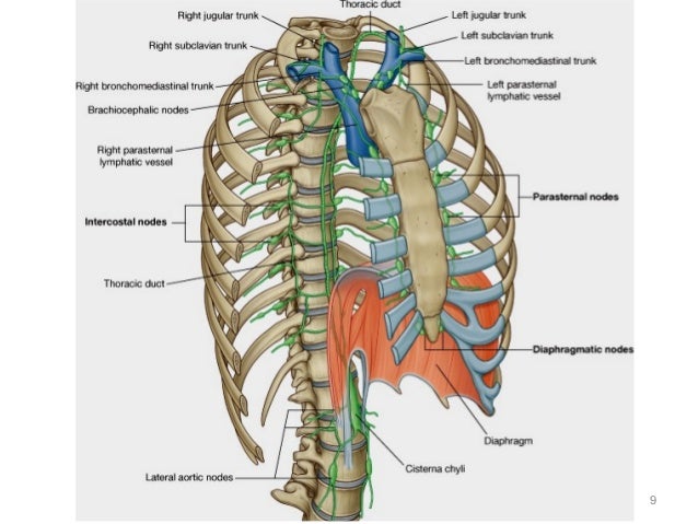
· Thoracic duct (slide 9) (SlideShare)
· Lymphatic trunks and ducts (antranik.org)

Where it is found
Related terms
- Thoracic aorta
- Azygos vein
- Esophagus
- Internal jugular vein
- Subclavian vein
- Brachiocephalic vein
- Abdominal aorta
References and sources
- Further reading: Thoracic duct, Wikipedia.
- Further reading: Cisterna chyli, Wikipedia.
- Federative International Programme for Anatomical Terminology (FIPAT). Terminologia Anatomica, 2nd ed. (TA2), 2019.