Latin: Vena iliaca communis
Overview
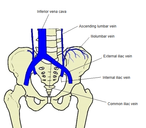
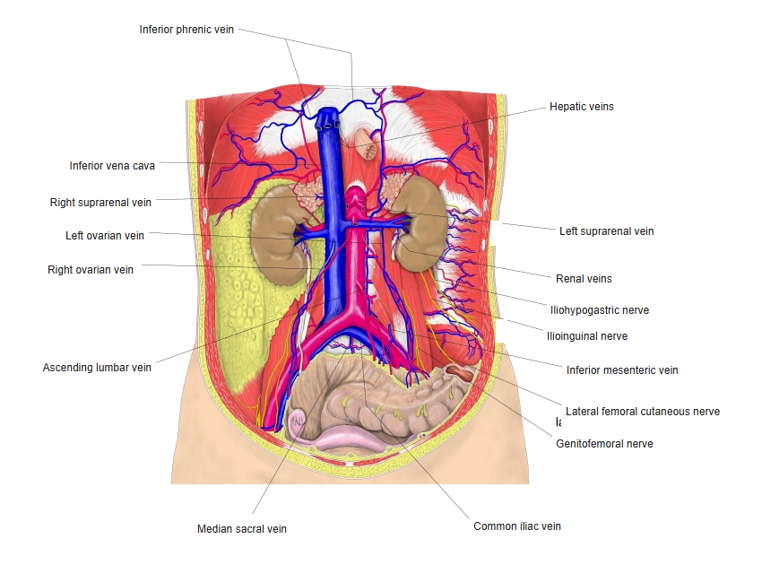
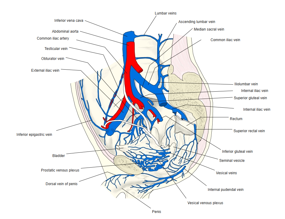
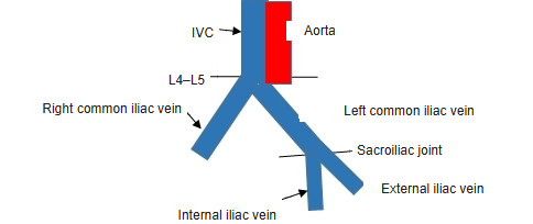
- Formed in front of the sacroiliac joint by the union of the external and internal iliac veins.
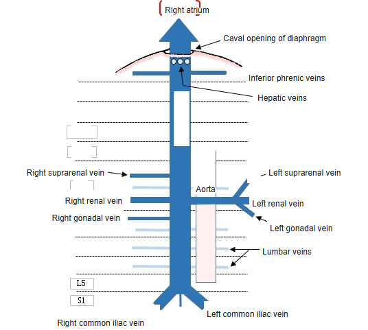
- The two common iliac veins join at the level of the 5th lumbar vertebra, to the right of the midline, to form the inferior vena cava.
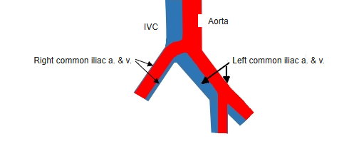
- The left vein is longer and more oblique; it passes behind the right common iliac artery.
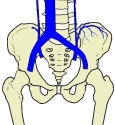
Iliolumbar vein 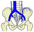
Median sacral vein and pelvic veins
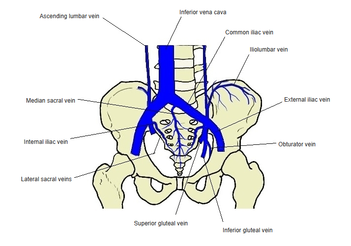
Tributaries and drainage
- Iliolumbar vein and sometimes the lateral sacral veins
- Left vein only: the median sacral vein
- The lower end of the ascending lumbar vein begins from it.
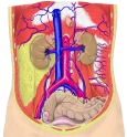
Veins of the posterior abdominal wall 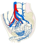
Veins of the male pelvis
Clinical notes
- May–Thurner syndrome: compression of the left common iliac vein by the right common iliac artery predisposes to left-sided deep vein thrombosis.
References and sources
- Further reading: Common iliac vein, Wikipedia.