Latin: Venae diploicae
Overview
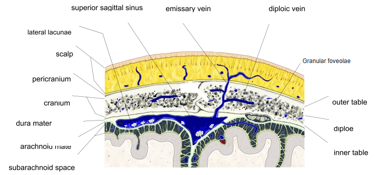
- Thin-walled, valveless veins in the diploë, the spongy bone between the outer and inner tables of the skull.
- Connect with the dural venous sinuses inside and with the veins of the scalp outside.
- Large irregular channels that appear after about the 2nd year of life.
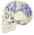
Diploic and emissary veins 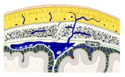
Layers of the scalp and skull
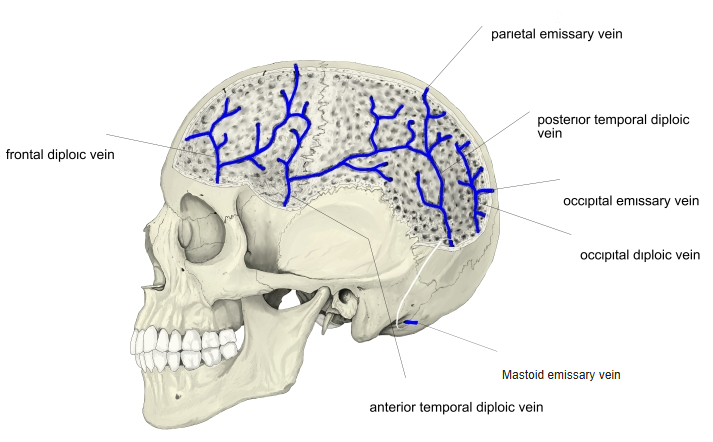
Main diploic veins
| Vein | Drains into |
|---|---|
| Frontal diploic vein | Supraorbital vein (outside) and superior sagittal sinus (inside) |
| Anterior temporal diploic vein | Sphenoparietal sinus or deep temporal veins |
| Posterior temporal diploic vein | Transverse sinus (through the mastoid foramen) |
| Occipital diploic vein | Occipital vein or transverse sinus |
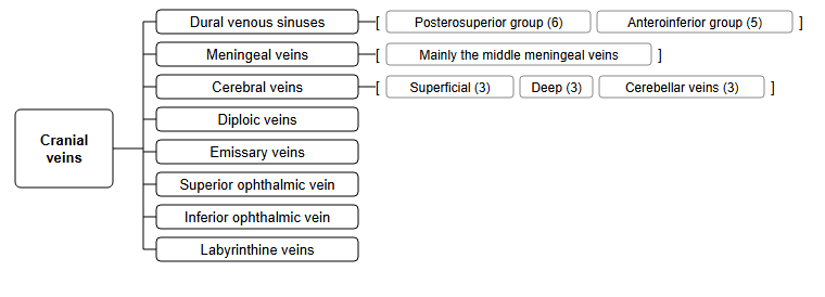
Place among the cranial veins
- The diploic veins are one of several groups of veins inside the skull that finally drain into the internal jugular vein.
Clinical notes
- Because they have no valves and connect the scalp with the sinuses, infection of the scalp can spread into the skull through these veins.
- Diploic channels can be seen on skull X-rays and should not be mistaken for fractures.
References and sources
- Further reading: Diploic veins, Wikipedia.