Calvaria (Latin: calvaria)
Also called the skullcap or the cranial vault
· The outer surface of the calvaria is covered by a strong periosteum called the pericranium.
Front: the front part, formed by part of the frontal bone.
Vertex: the middle part, formed by the parietal bones.
Back: the back part, formed by part of the occipital bone (the occipital squama).
Temporal plane: the part on each side below the temporal lines, where temporalis arises; formed by the parietal bone and part of the temporal bone (its squamous part).
Note: some sources do not include the temporal bone in the calvaria.
| 1 | Outer table | The outermost layer, of thick compact bone. |
| 2 | Diploe | The middle layer, of spongy bone with red marrow. It contains the diploic canals, in which veins run (the diploic veins, which communicate with the emissary veins). |
| 3 | Inner table | The innermost layer, of thin, more brittle compact bone, facing the dura mater. On its inner surface are: 1. Digital impressions: made by the gyri of the brain 2. Cerebral juga: ridges matching the sulci 3. Grooves for arteries and veins: made by the vessels, such as the branches of the middle meningeal artery, and the groove for the superior sagittal sinus in the midline 4. Granular foveolae: small pits that hold the arachnoid granulations of the arachnoid mater Digital impressions and juga are much fainter on the calvaria than on the cranial base. |
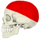 Calvaria (lateral view)
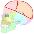 Bones of the calvaria
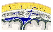 Layers of the scalp, calvaria and meninges
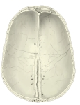 Calvaria (internal surface)
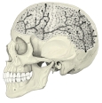 Diploe and diploic canals
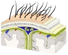 Scalp, skull and meninges (coronal)
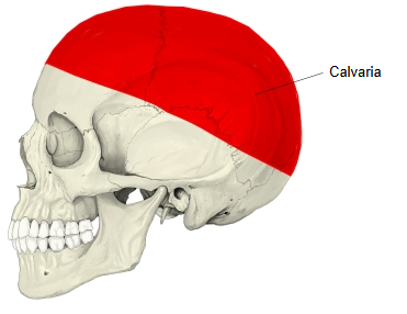
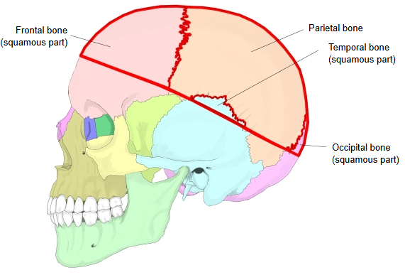
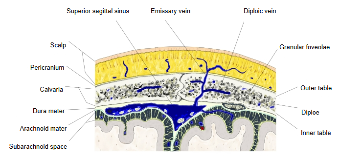
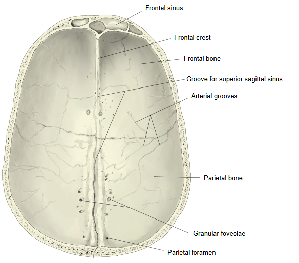
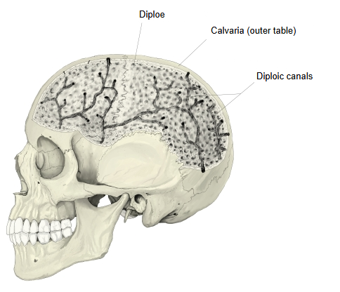
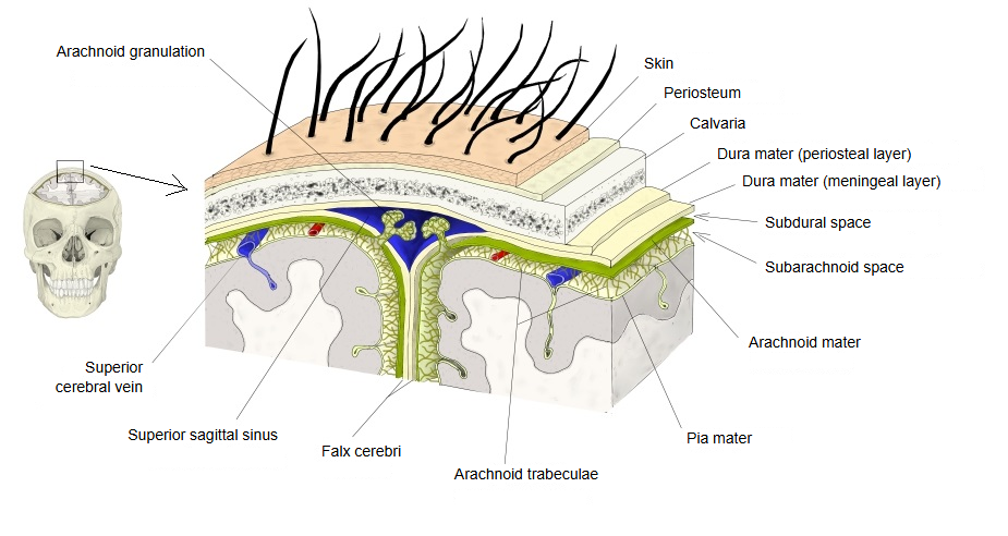
These bones are flat bones with an outer and an inner table of compact bone and, between them, the diploe of spongy bone. The diploe contains the diploic canals for veins, which open on the outer or inner table. The bones develop directly in membrane (intramembranous ossification) and are joined by the coronal, sagittal and lambdoid sutures; in the newborn the sutures are still wide and the fontanelles can be felt, and early closure of a suture is called craniosynostosis. The outer surface of the calvaria is smooth, without marked bumps, and oval when seen from above. The slight bulges of the frontal and parietal bones are the frontal tuber and the parietal tuber; on each side the superior and inferior temporal lines can be seen.
As noted above, some sources do not include the temporal bone in the calvaria.
For example, the calvaria (skullcap) is often described as made up of the upper parts of the frontal bone, the occipital bone and the two parietal bones only. The Latin word is calvaria (plural calvariae), though 'calvarium' is also widely used.
Reference websites
[Wikipedia]
· Calvaria (skull), Wikipedia.
[Websites with illustrations]
· Skull, lateral view, with the bones of the calvaria shaded (photograph) (academic.ru (medical dictionary))

· Skull, lateral view, with the calvaria shaded (drawing) (The Free Dictionary (Medical Dictionary))

Where it is found
Related terms
- Frontal bone
- Parietal bone
- Occipital bone
- Diploic veins
- Granular foveolae
- Sagittal suture
- Temporal line
References and sources
- Further reading: Calvaria (skull), Wikipedia.
- Federative International Programme for Anatomical Terminology (FIPAT). Terminologia Anatomica, 2nd ed. (TA2), 2019.