Latin: Arteria carotis externa
Overview
- One of the two terminal branches of the common carotid artery; the other is the internal carotid artery.
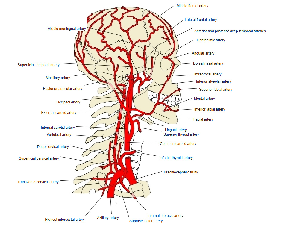
- Supplies most structures of the head and neck outside the skull: the face, scalp, tongue, thyroid gland, pharynx and jaws (with a branch to the dura mater).
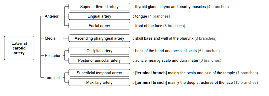
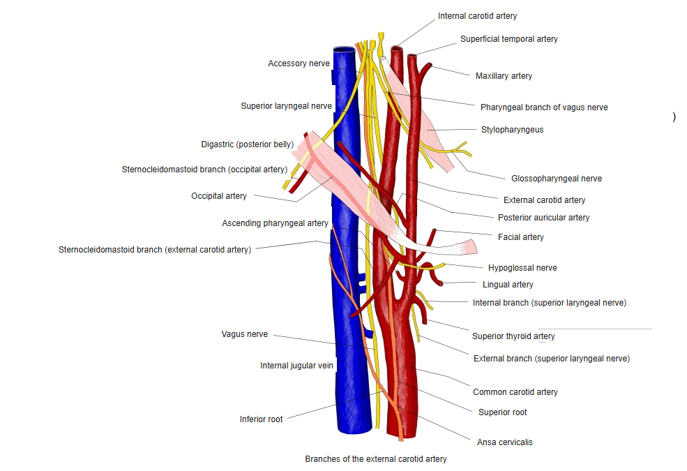
- Has eight branches, usually grouped by direction: anterior (3), medial (1), posterior (2) and terminal (2).
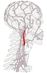
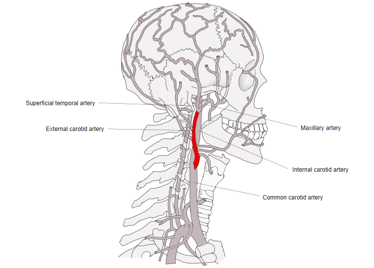
External carotid artery (lateral view) 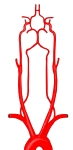
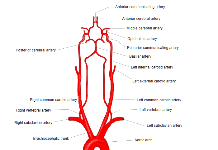
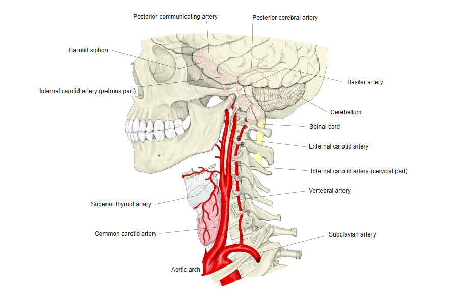
Carotid and vertebral arteries (front view)

Origin
- Arises at the bifurcation of the common carotid artery, usually at the level of the upper border of the thyroid cartilage (about C3–C4).
- At first it lies in front of and medial to the internal carotid artery, then it moves to its lateral side as it ascends.
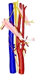
Common, external and internal carotid arteries
Course
- Ascends in the carotid triangle, covered by the sternocleidomastoid, then passes deep to the posterior belly of the digastric and the stylohyoid.
- Enters the parotid gland, and ends behind the neck of the mandible by dividing into the superficial temporal and maxillary arteries.
Branches of the external carotid artery (front view) 
Arteries of the head and neck
Branches
| Group | Branch | Main area supplied |
|---|---|---|
| Anterior | Superior thyroid artery | Thyroid gland, larynx and nearby muscles |
| Anterior | Lingual artery | Tongue and floor of the mouth |
| Anterior | Facial artery | Front of the face, palatine tonsil |
| Medial | Ascending pharyngeal artery | Pharynx, skull base, middle ear |
| Posterior | Occipital artery | Back of the scalp and neck muscles |
| Posterior | Posterior auricular artery | Auricle, scalp behind the ear, middle ear |
| Terminal | Superficial temporal artery | Scalp and skin of the temple |
| Terminal | Maxillary artery | Deep structures of the face |
Clinical notes
- The pulse of the superficial temporal artery (in front of the ear) and the facial artery (at the lower border of the mandible) can be felt.
- Ligation of the external carotid artery is relatively safe because its branches anastomose widely with those of the opposite side.
References and sources
- Further reading: External carotid artery, Wikipedia.