Carotid triangle (Latin: trigonum caroticum)
Also called the superior carotid triangle
- Boundaries and floor
- Arteries
- Veins
- Nerves
- Other contents
- Reference websites
- (Wikipedia, websites with illustrations)
- Where it is found
- Related terms
There is one on each side, in the upper front of the neck, just in front of the upper part of sternocleidomastoid. It is part of the anterior triangle, and the carotid arteries lie close to the surface here.
For more detail, see Wikipedia below.
The middle of the triangle corresponds to the point where the common carotid artery divides into the internal and external carotid arteries, at about the level of the upper border of the thyroid cartilage (the carotid bifurcation); a strong pulse can be felt here. The carotid sinus and the carotid body lie at the bifurcation. In the triangle the external carotid artery gives off the superior thyroid, ascending pharyngeal, lingual and facial arteries. Because the main vessels and nerves of the neck are fairly superficial here, it is a common surgical approach to them.
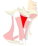 Carotid triangle (right side)
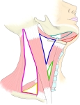 Triangles of the neck
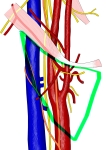 Vessels and nerves in the carotid triangle
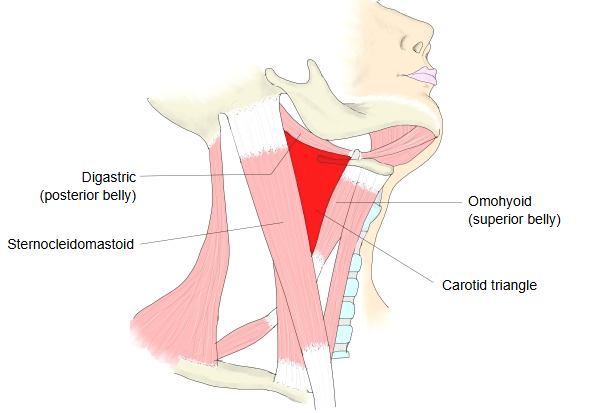
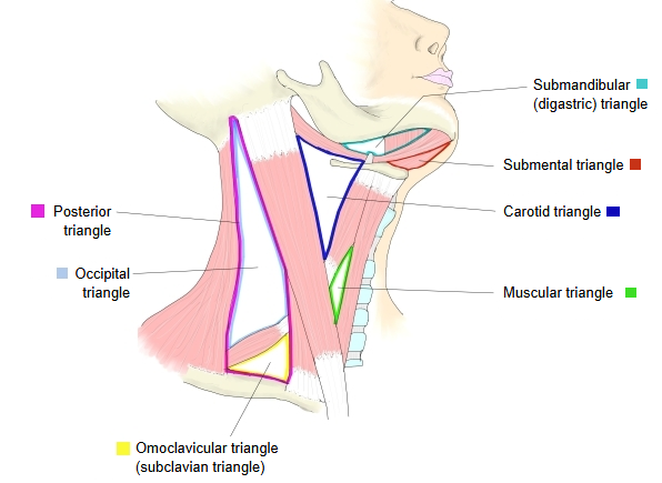
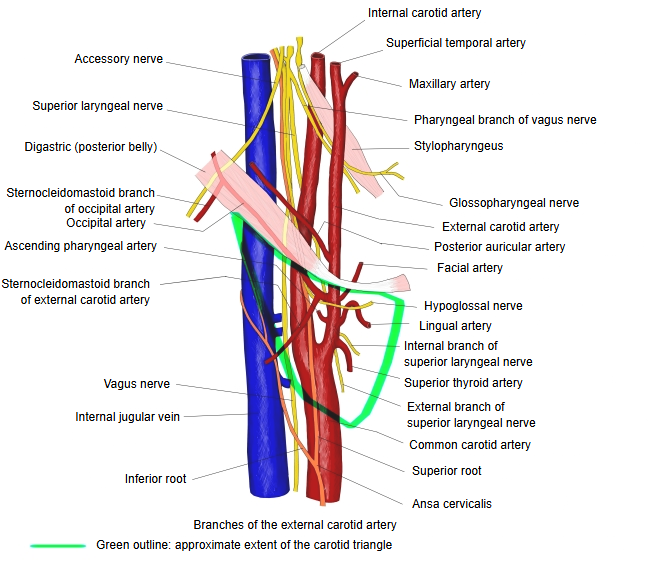
Below is a simple outline of the triangular depressions of the neck (it may not include every one).
depressions
of the neck
- Anterior triangle : sternocleidomastoid (anterior border), midline of the neck, body of mandible (lower border)
- Submandibular triangle : digastric (posterior and anterior bellies), body of mandible
- Submental triangle : anterior bellies of the left and right digastric, hyoid bone
- Carotid triangle : sternocleidomastoid, digastric (posterior belly), omohyoid (superior belly)
- Muscular triangle : sternocleidomastoid (anterior border), omohyoid, midline of the neck = omotracheal triangle
- Posterior triangle : sternocleidomastoid (posterior border), trapezius (anterior border), clavicle (middle third) = lateral cervical region
- Occipital triangle : trapezius (anterior border), sternocleidomastoid (posterior border), omohyoid (inferior belly)
- Omoclavicular triangle : omohyoid (inferior belly), sternocleidomastoid (posterior border), clavicle (middle third)
Boundaries and floor
The triangle is bounded:
· behind, by the anterior border of sternocleidomastoid;
· in front and below, by the superior belly of omohyoid;
· above, by the posterior belly of digastric.
Its roof is formed by the skin, the superficial fascia with the platysma, and the investing layer of the deep cervical fascia; branches of the facial nerve and of the cutaneous nerves of the neck run in it. Its floor is formed by parts of:
· the thyrohyoid membrane;
· hyoglossus;
· the middle and inferior pharyngeal constrictors.
Arteries
The end of the common carotid artery and the beginnings of the internal and external carotid arteries lie here side by side, the external carotid in front of the internal. Branches of the external carotid artery met in this space (some may arise just outside the triangle):
· the superior thyroid artery, running forwards and downwards;
· the lingual artery, running forwards;
· the facial artery, running forwards and upwards;
· the occipital artery, running backwards and upwards;
· the ascending pharyngeal artery, running straight up on the medial side of the internal carotid artery.
Veins
The veins in the triangle are:
· the internal jugular vein, on the lateral side of the common and internal carotid arteries;
and veins that accompany the branches of the external carotid artery and drain into the internal jugular vein:
· the superior thyroid vein;
· the lingual veins;
· the common facial vein;
· the pharyngeal veins, which accompany the ascending pharyngeal artery and usually end in the internal jugular vein;
· and sometimes the occipital vein.
Nerves
The vagus nerve lies within the carotid sheath, between and behind the artery and the vein; the sympathetic trunk lies behind the sheath. On the sheath lies the superior root of the ansa cervicalis of the cervical plexus. The hypoglossal nerve crosses the internal and external carotid arteries, curving round the origin of the occipital artery. The accessory nerve runs for a short distance near the upper corner, lateral to the vessels, before it enters sternocleidomastoid. Medial to the external carotid artery, just below the hyoid bone, lies the internal branch of the superior laryngeal nerve, and lower down its external branch.
Other contents
Deep cervical lymph nodes lie along the internal jugular vein, and the upper part of the larynx and the lower part of the pharynx lie towards the front of the triangle.
Reference websites
[Wikipedia]
· Carotid triangle, Wikipedia.
· Anterior triangle of the neck, Wikipedia.
[Websites with illustrations]
· Anterior triangle of the neck: boundaries, contents and subdivisions (TeachMeAnatomy)
· Muscles of the neck with the carotid triangle marked (Wikimedia Commons)

· Carotid triangle (illustration) (earthslab.com)
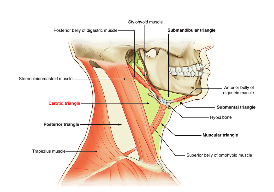
· Contents of the carotid triangle (illustration) (anatomyqa.com)

Where it is found
Related terms
- Carotid sheath
- Carotid sinus
- Common carotid artery
- External carotid artery
- Internal carotid artery
- Sternocleidomastoid
- Omohyoid
References and sources
- Further reading: Carotid triangle, Wikipedia.
- Further reading: Anterior triangle of the neck, Wikipedia.
- Federative International Programme for Anatomical Terminology (FIPAT). Terminologia Anatomica, 2nd ed. (TA2), 2019.