Latin: Venae renales
Overview
- Large, short veins that drain each kidney into the inferior vena cava, at about the level of L1–L2.
- Formed in the renal sinus by several veins; leave the hilum in front of the renal artery and the renal pelvis.
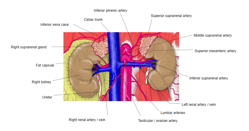
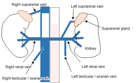
- The left renal vein is about three times longer than the right: it crosses in front of the aorta just below the origin of the superior mesenteric artery.
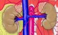
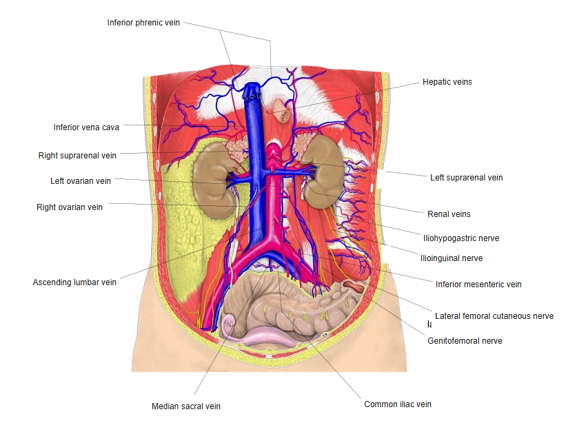
Kidneys and suprarenal glands with their vessels 
Venae cavae and their tributaries
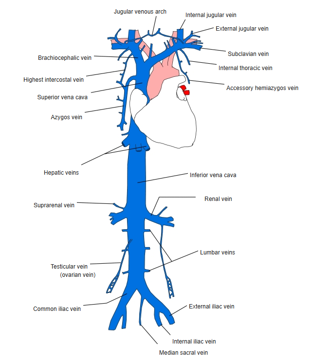
Tributaries and drainage
- Left renal vein receives the left suprarenal vein, the left testicular or ovarian vein, the left inferior phrenic vein and often a lumbar vein.
- Right renal vein usually receives no tributaries (the right gonadal and suprarenal veins end in the IVC).
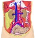
Veins of the posterior abdominal wall
Clinical notes
- Nutcracker syndrome: compression of the left renal vein between the aorta and the superior mesenteric artery can cause blood in the urine and a left varicocele.
- Variants: a left renal vein passing behind the aorta (retroaortic) or around it (circumaortic).
- The longer left vein is preferred in living-donor kidney transplantation.
References and sources
- Further reading: Renal veins, Wikipedia.