Latin: Vena suprarenalis
Overview
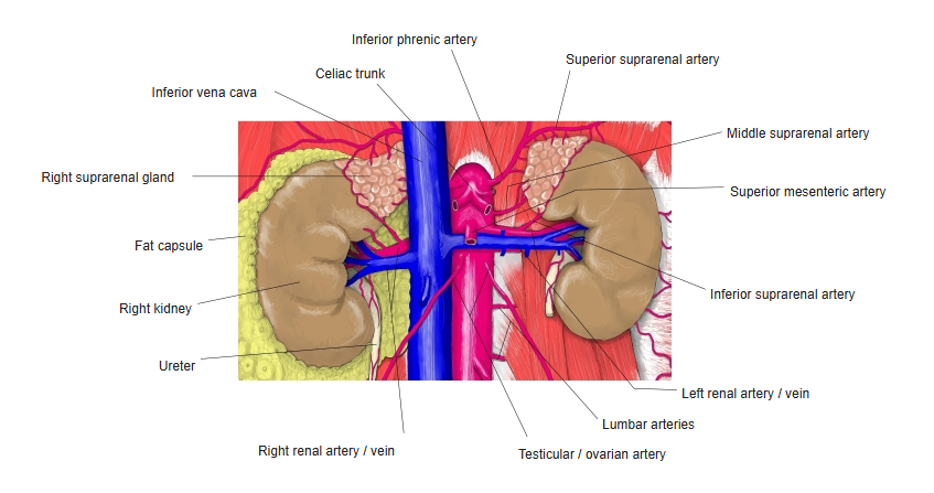
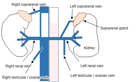
- Each suprarenal (adrenal) gland is drained by a single vein leaving its hilum.
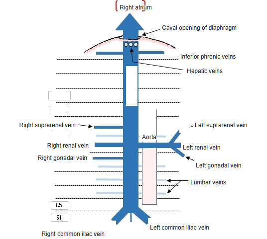
- The right vein is very short and ends directly in the back of the inferior vena cava.
- The left vein runs down and ends in the left renal vein, often joined by the left inferior phrenic vein.
Kidneys and suprarenal glands with their vessels 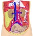
Veins of the posterior abdominal wall
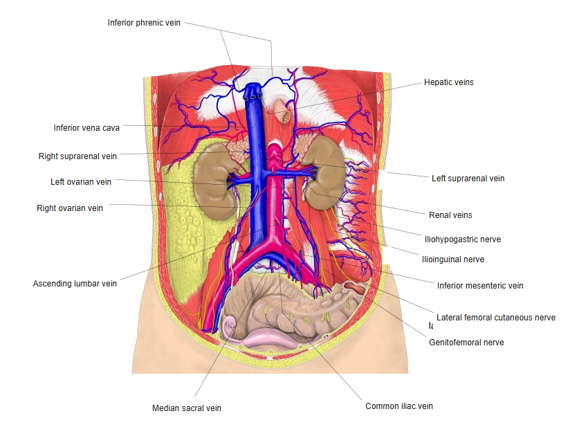
Tributaries and drainage
- The gland has many arteries (superior, middle and inferior suprarenal) but only one main vein.
Clinical notes
- Adrenal venous sampling (for primary aldosteronism) needs a catheter in each suprarenal vein; the short right vein is the harder one to enter.
References and sources
- Further reading: Suprarenal vein, Wikipedia.