Kidney (Latin: ren; nephros)
- Overview (shape and size)
- Location (surrounding organs)
- Structure (renal cortex, renal medulla, calyces and renal pelvis, nephron)
- Kidney and its coverings
- Function
- Vessels and nerves
- Renal segments
- Related terms
Overview
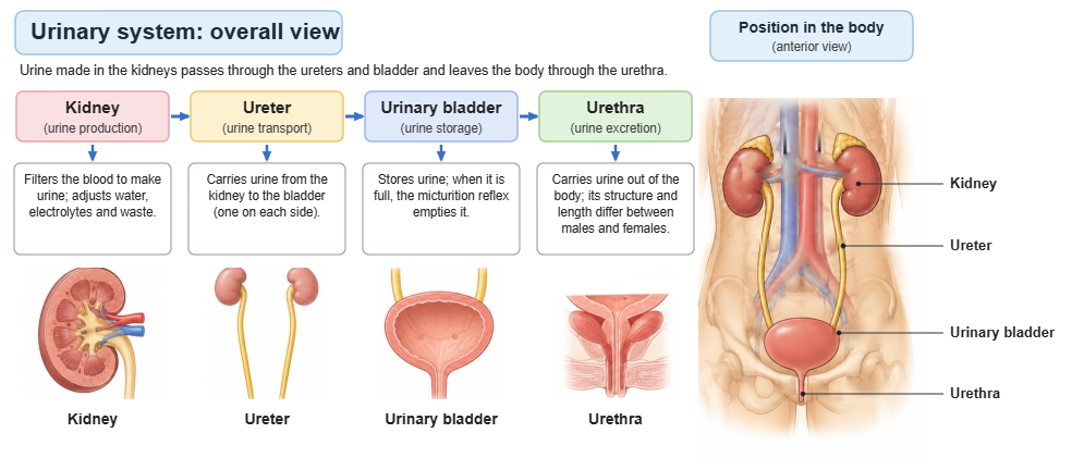
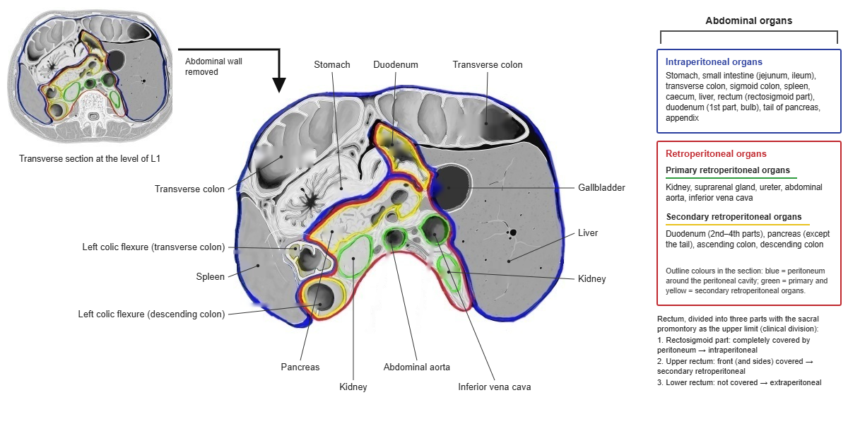
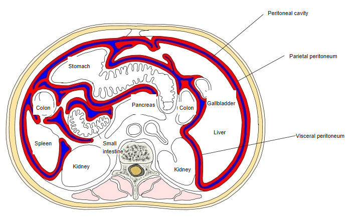
 Intraperitoneal / retroperitoneal organs
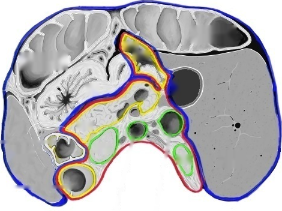
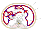 Peritoneum in a transverse section
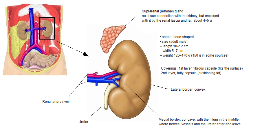
[Shape]
Adult male: length 10–12 cm, width 5–7 cm, thickness about 3 cm; weight about 120–170 g.
Location
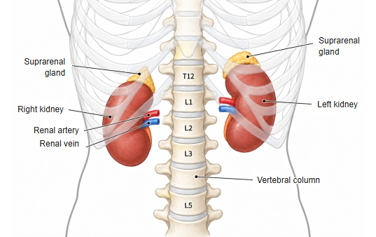
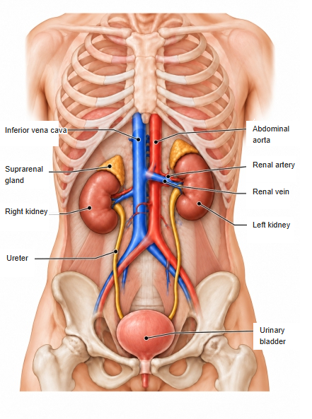
The kidney is a retroperitoneal organ lying behind the abdominal cavity, on each side of the vertebral column.
It extends roughly from the 12th thoracic vertebra (T12) to the 3rd lumbar vertebra (L3). Because the large liver lies on the right, the right kidney is usually slightly lower than the left.
The suprarenal gland (adrenal gland) sits near the upper end of each kidney. The hilum faces medially and forward, and the kidneys move a few centimetres up and down with breathing.
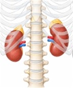 Vertebral column and kidneys
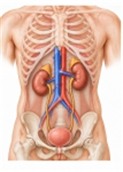 Urinary organs
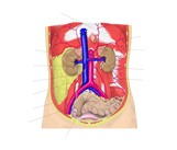 Position of the kidneys
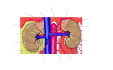 Kidneys, suprarenal glands and their arteries
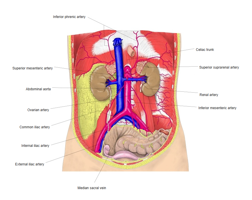
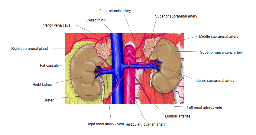
[Surrounding organs]
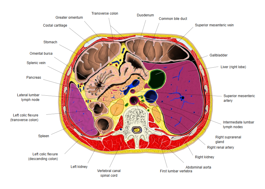
The kidneys lie behind the peritoneum, and the organs in front of them differ between the right and left sides.
[Right kidney]
In front of the right kidney lie mainly the liver, the duodenum (descending part) and the ascending colon (right colic flexure).
[Left kidney]
In front of the left kidney lie mainly the stomach, spleen, pancreas, jejunum and descending colon (left colic flexure).
[Behind]
Behind, both kidneys lie on the diaphragm, psoas major and quadratus lumborum (and transversus abdominis). The 12th rib crosses behind the right kidney, the 11th and 12th ribs behind the left.
Structure
· The renal parenchyma makes up about 85–90% of the kidney and the renal sinus about 10–15%.
* Figures vary between sources; some give 70–80% for the parenchyma.
- Renal parenchyma : the part that makes urine and controls its composition; divided into the renal cortex and the renal medulla.
About 85–90% of the kidney. - Renal sinus : an irregular space enclosed by the parenchyma, holding fat, blood vessels and the urinary passages.
About 10–15% of the kidney.
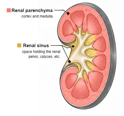
The renal parenchyma, the working part of the kidney, is divided into the renal cortex and the renal medulla.
- Renal cortex
- Renal corpuscle : glomerulus and Bowman's capsule.
- Proximal tubule : takes back water and nutrients from the filtrate.
- Distal tubule : adjusts electrolytes and water.
- Cortical collecting duct : collects urine from several distal tubules.
- Renal medulla
- Renal pyramid : the cone-shaped tissue that forms the medulla; it contains:
- Loop of Henle : part of the countercurrent system that concentrates urine.
- Medullary collecting duct : carries urine to the renal papilla.
- Papillary duct : opens at the tip of the papilla and drains urine into a minor calyx.
- Renal pyramid : the cone-shaped tissue that forms the medulla; it contains:
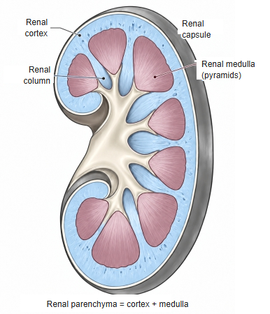
The kidney can also be divided into the renal parenchyma (renal cortex and renal medulla), which makes urine, and the other parts (renal sinus, renal hilum, capsule and other coverings).
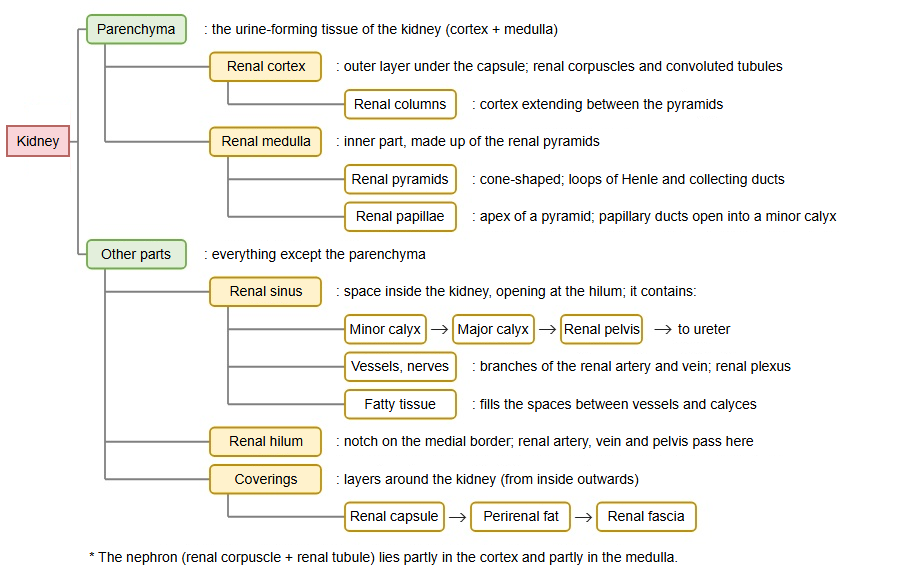
Calyces and renal pelvis: the cortex forms the outer layer and extends between the 8–18 renal pyramids as the renal columns. Urine leaves each renal papilla into a minor calyx; minor calyces join to form 2–3 major calyces, which open into the funnel-shaped renal pelvis. The pelvis continues as the ureter at the hilum.
Nephron: the functional unit of the kidney (about one million in each kidney): renal corpuscle → proximal tubule → loop of Henle → distal tubule. Its tubular part is the renal tubule. Several nephrons drain into one collecting duct, which runs through the medulla to a papilla.
Kidney and its coverings
| Name | Features | |
|---|---|---|
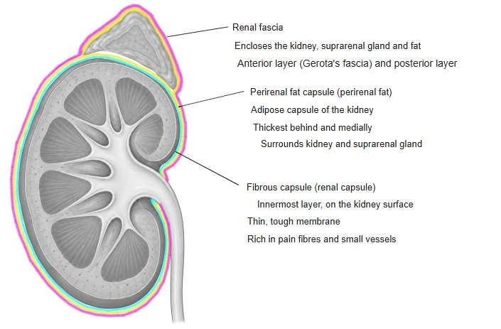
| 1 | (= fibrous capsule) | Innermost thin, tough fibrous membrane that directly covers the kidney surface. Composition: mostly dense, regularly arranged collagen fibres (types I and III), with a few elastic and smooth muscle fibres. The term "fibrous capsule" is also used for the liver, spleen, testis and ovary. |
| 2 | (Perirenal) (= perirenal fat) | Fat layer outside the fibrous capsule, best developed behind and medially; it also encloses the suprarenal gland. |
| 3 | (anterior layer = Gerota's fascia) | Fibrous sheet that encloses the kidney, suprarenal gland and surrounding fat together; it has two layers (anterior and posterior). Composition: mainly collagen fibres (types I and III), loosely mixed with many elastic fibres. |
Outside the renal fascia lies the pararenal fat, mainly behind the kidney.
Function
[1. Urine production and excretion of waste]
Filters the blood and removes urea, creatinine, uric acid and other waste products in the urine.
[2. Body-fluid volume]
Adjusts how much water is reabsorbed or excreted, keeping body-fluid and blood volume within range.
[3. Electrolytes]
Regulates the reabsorption and excretion of sodium, potassium, calcium and other ions, keeping their concentrations within range.
[4. Acid–base balance]
Excretes hydrogen ions and reabsorbs bicarbonate, keeping the pH of the blood within range.
[5. Blood pressure]
The juxtaglomerular cells release renin, which acts through the renin–angiotensin–aldosterone system (RAAS) on blood pressure and body-fluid volume.
[6. Red blood cell production]
The kidney produces erythropoietin (EPO), which stimulates the bone marrow to make red blood cells.
[7. Vitamin D activation]
The kidney converts vitamin D to its active form (calcitriol), which is important for calcium, phosphate and bone metabolism.
Vessels and nerves
[Arteries]
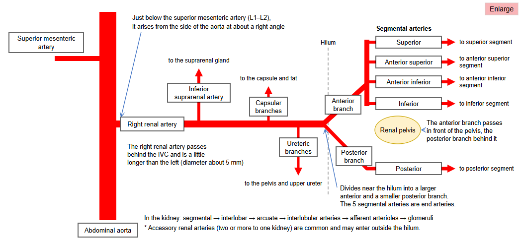
Each renal artery arises from the abdominal aorta (at about L1–L2) and enters the kidney at the hilum.
The main arterial route inside the kidney:
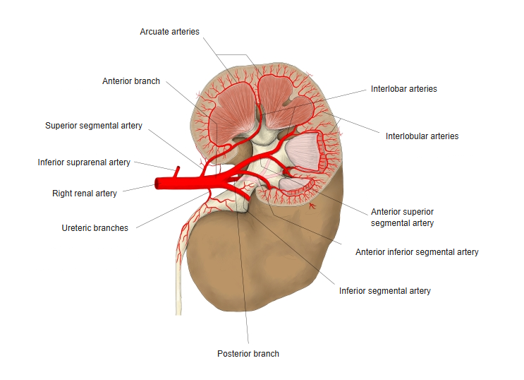
Abdominal aorta → renal artery → segmental arteries → interlobar arteries → arcuate arteries → interlobular (cortical radiate) arteries → afferent arteriole → glomerulus
Blood leaving the glomerulus passes into the efferent arteriole.
[Veins]
Venous blood from the kidney finally collects in the renal vein, which drains into the inferior vena cava.
The left renal vein is longer than the right and crosses in front of the abdominal aorta to reach the inferior vena cava.
[Lymphatics]
Lymph vessels run mainly along the renal vessels to the lumbar (lateral aortic) lymph nodes.
[Nerves]
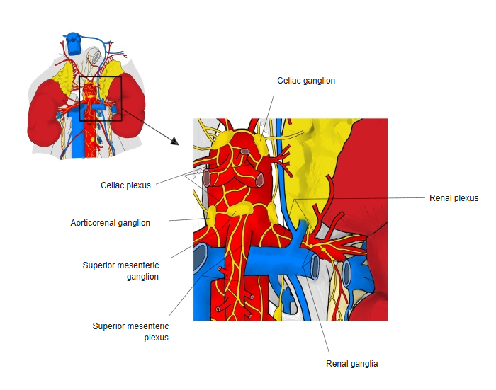
Autonomic fibres reach the kidney through the renal plexus.
Sympathetic fibres in particular constrict the renal vessels and stimulate renin release.
Visceral afferent fibres from the kidney and ureter carry pain, so pain from these organs may be felt in the back and flank (often spreading toward the groin).
 Renal plexus
Renal segments
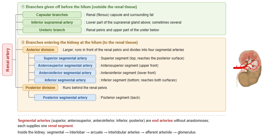
The renal segments divide the kidney according to the territory supplied by each segmental artery of the renal artery. There are five: superior, anterosuperior, anteroinferior, inferior and posterior.
Segmental arteries are end arteries without anastomoses, so if one is blocked only its own segment loses its blood supply (renal infarction). The segments therefore matter in operations that deal with the vessels segment by segment, such as partial nephrectomy.
The segments do not match the renal lobes (one renal pyramid with the cortex over it).
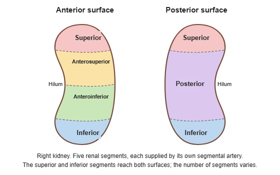
[The five segments and their arteries]
| Segment | Artery | Position |
|---|---|---|
| Superior | Superior segmental artery (anterior division) | Top of the kidney; extends from the anterior to the posterior surface. |
| Anterosuperior | Anterosuperior segmental artery (anterior division) | Upper part of the anterior surface. |
| Anteroinferior | Anteroinferior segmental artery (anterior division) | Lower part of the anterior surface. |
| Inferior | Inferior segmental artery (anterior division) | Bottom of the kidney; reaches both surfaces. |
| Posterior | Posterior segmental artery (posterior division) | Most of the posterior surface. |
Near the hilum the renal artery divides into an anterior division and a posterior division. The anterior division passes in front of the renal pelvis and gives four segmental arteries; the posterior division passes behind the pelvis and becomes the posterior segmental artery.
[Avascular plane]
Between the territories of the two divisions lies a plane with few vessels (Brödel's line), slightly behind the lateral border. It is used as the route for reaching the renal pelvis through the skin (e.g. percutaneous nephrostomy).
[Variation in the number of segments]
Five segments is the standard model; the number of segments and the branching of the segmental arteries vary widely. Corrosion-cast studies have reported 5–9 segments (about 7 on average).
Related terms
Main anatomical terms related to the kidney, in alphabetical order.
References and sources
- Further reading: Kidney, Wikipedia.